Review
. 2021 Mar 13;10(3):639.
doi: 10.3390/cells10030639.

Overview on the Different Patterns of Tumor Vascularization

Affiliations
Review

Overview on the Different Patterns of Tumor Vascularization

Domenico Ribatti et al. Cells. .

Abstract

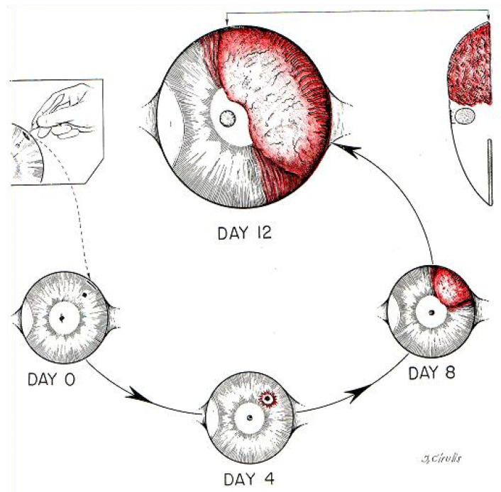
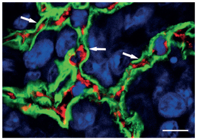
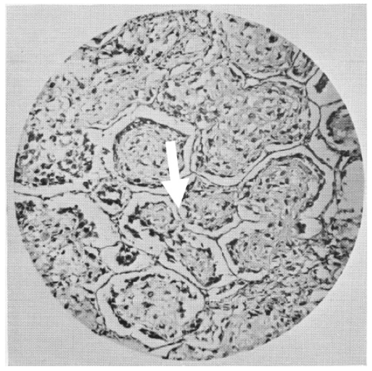
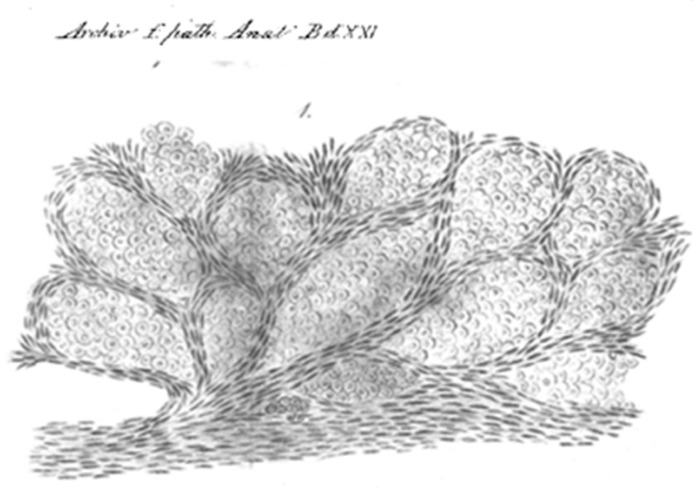
Angiogenesis is a crucial event in the physiological processes of embryogenesis and wound healing. During malignant transformation, dysregulation of angiogenesis leads to the formation of a vascular network of tumor-associated capillaries promoting survival and proliferation of the tumor cells. Starting with the hypothesis formulated by Judah Folkman that tumor growth is angiogenesis-dependent, this area of research has a solid scientific foundation and inhibition of angiogenesis is a major area of therapeutic development for the treatment of cancer. Over this period numerous authors published data of vascularization of tumors, which attributed the cause of neo-vascularization to various factors including inflammation, release of angiogenic cytokines, vasodilatation, and increased tumor metabolism. More recently, it has been demonstrated that tumor vasculature is not necessarily derived by endothelial cell proliferation and sprouting of new capillaries, but alternative vascularization mechanisms have been described, namely vascular co-option and vasculogenic mimicry. In this article, we have analyzed the mechanisms involved in tumor vascularization in association with classical angiogenesis, including post-natal vasculogenesis, intussusceptive microvascular growth, vascular co-option, and vasculogenic mimicry. We have also discussed the role of these alternative mechanism in resistance to anti-angiogenic therapy and potential therapeutic approaches to overcome resistance.

Keywords: angiogenesis; tumor growth; vascular co-option; vasculogenic mimicry.


Conflict of interest statement

The authors declare no conflict of interest.

Figures

Figure 1
Figure 1
The patterns of development of two simultaneous implants of Brown-Pearce tumor in the rabbit eye. Tumors suspended in the aqueous fluid of the anterior chamber of the rabbit eye and observed for a period up to 6 weeks remain viable, avascular, of limited size (less than 1 mm3) and contain a population of viable and mitotically active tumor cells. After implantation contiguous to the iris, which has abundant blood vessels, the tumors induced neovascularization and grow rapidly, reaching 16,000 times the original size within two weeks. The anterior chamber implant remains avascular, while the iris implant vascularizes and grow progressively. (Reproduced from [31]).
Figure 2
Figure 2
Preservation of alveolar architecture in the peripheral regions of lung metastases that is indicative of vessel co-option. Experimental lung metastases are generated by injecting HT1080 cells intravenously into SCID mice. After extravasation and forming small interstitial colonies, tumor cells enter the alveolar air spaces. This high-power confocal micrograph of the periphery of an HT1080 lung metastasis has been stained for podoplanin (green), CD31 (red), and with TOTO-3 (blue), which highlights the tumor mass. Note that intact alveolar walls with regular layering (pneumocyte–capillary–pneumocyte) are present (arrows). Scale bar = 10 μm. (Reproduced from [52]).
Figure 3
Figure 3
A non-angiogenic lung metastases from an osteosarcoma presented by Rupert A. Wills in his 1934 book The Spread of the Tumours in the Human Body. In the words of the author: “Sections of a pulmonary metastasis showing alveoli occupied by plugs of growth resembling osteoid tissue. Note the strands of growth connecting the alveolar plugs trough the septal pores.” (arrow).
Figure 4
Figure 4
Camera Lucida drawing from a Johannes Erichsen paper of 1861 [58] is the oldest reproduction, in patients with tumors in the lung, of the neoplastic cells occupying the alveolar spaces. No new vessels can be seen while the alveolar septa containing the pre-existing capillaries, produce the “chicken wire” appearance. In the word of the author: “Cross section through a carcinoma nodule of the lungs: the alveoli are shaped by cancerous masses confined between the elastic fibers of the lung tissue”.

References

    1. Hall A.P. The role of angiogenesis in cancer. Comp. Clin. Pathol. 2005;13:95–99. doi: 10.1007/s00580-004-0533-3. - DOI
    1. Lenzi P., Bocci G., Natale G. John Hunter and the origin of the term “angiogenesis”. Angiogenesis. 2016;19:255–256. doi: 10.1007/s10456-016-9496-7. - DOI - PubMed
    1. Natale G., Bocci G., Lenzi P. Looking for the Word “Angiogenesis” in the History of Health Sciences: From Ancient Times to the First Decades of the Twentieth Century. World J. Surg. 2016;41:1625–1634. doi: 10.1007/s00268-016-3680-1. - DOI - PubMed
    1. Hunter J., Owen R. Essays and Observations on Natural History, Anatomy, Physiology, Psychology, and Geology. John van Voorst; London, UK: 1861. - DOI
    1. Flint J.M. The angiology, angiogenesis, and organogenesis of the submaxillary gland. Am. J. Anat. 1903;2:417–444. doi: 10.1002/aja.1000020402. - DOI

MeSH terms

LinkOut - more resources