

Mg,Si-Co-Substituted Hydroxyapatite/Alginate Composite Beads Loaded with Raloxifene for Potential Use in Bone Tissue Regeneration
- PMID: 33805785
- PMCID: PMC7999305
- DOI: 10.3390/ijms22062933
Mg,Si-Co-Substituted Hydroxyapatite/Alginate Composite Beads Loaded with Raloxifene for Potential Use in Bone Tissue Regeneration
Abstract
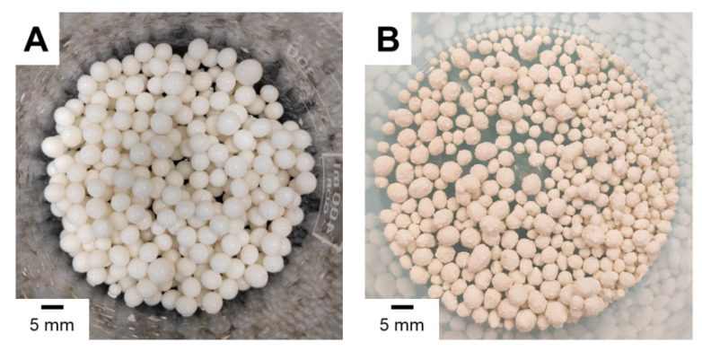
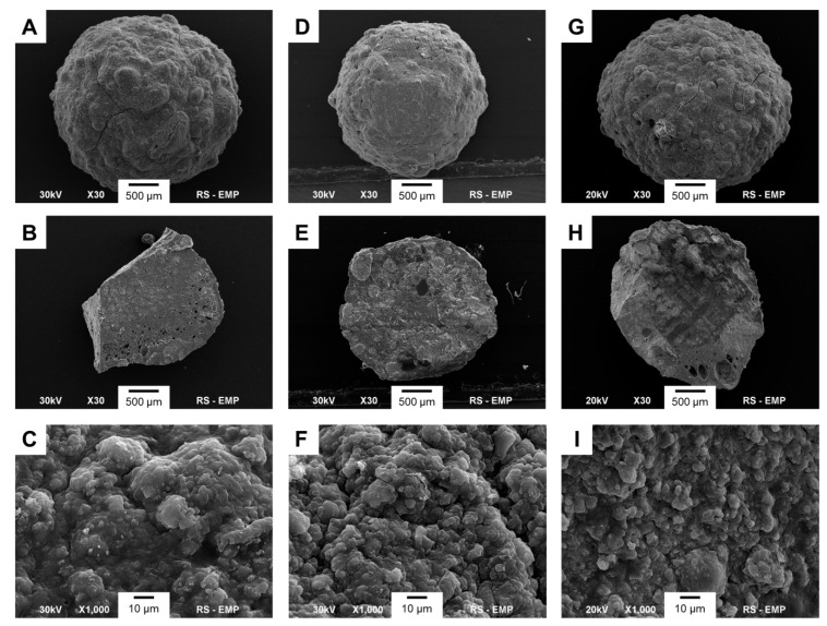
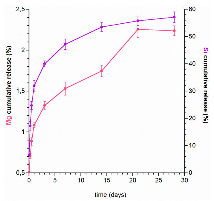
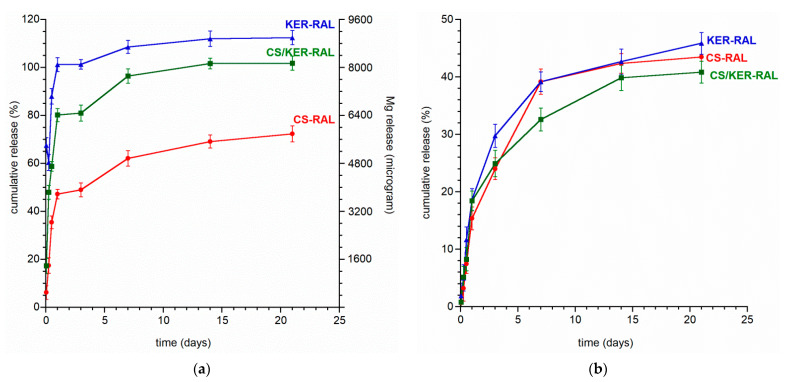
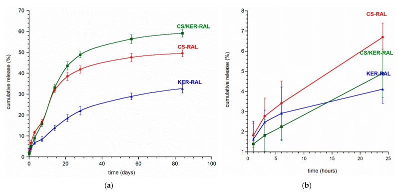
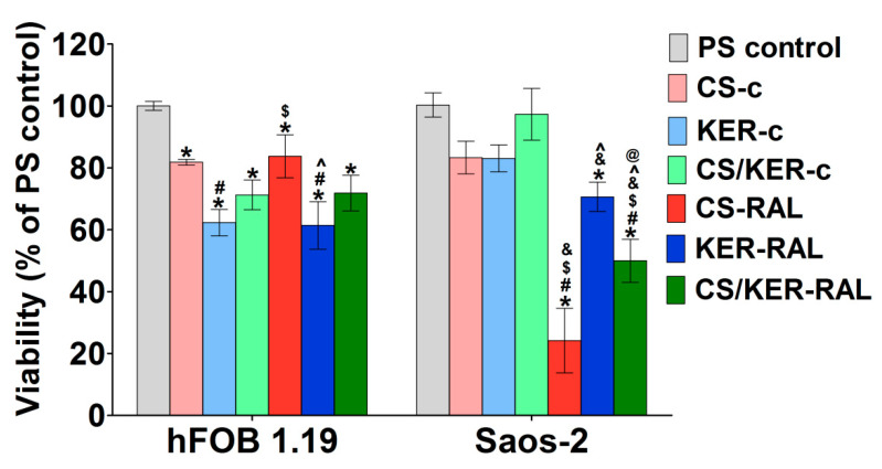
Osteoporosis is a worldwide chronic disease characterized by increasing bone fragility and fracture likelihood. In the treatment of bone defects, materials based on calcium phosphates (CaPs) are used due to their high resemblance to bone mineral, their non-toxicity, and their affinity to ionic modifications and increasing osteogenic properties. Moreover, CaPs, especially hydroxyapatite (HA), can be successfully used as a vehicle for local drug delivery. Therefore, the aim of this work was to fabricate hydroxyapatite-based composite beads for potential use as local carriers for raloxifene. HA powder, modified with magnesium and silicon ions (Mg,Si-HA) (both of which play beneficial roles in bone formation), was used to prepare composite beads. As an organic matrix, sodium alginate with chondroitin sulphate and/or keratin was applied. Cross-linking of beads containing raloxifene hydrochloride (RAL) was carried out with Mg ions in order to additionally increase the concentration of this element on the material surface. The morphology and porosity of three different types of beads obtained in this work were characterized by scanning electron microscopy (SEM) and mercury intrusion porosimetry, respectively. The Mg and Si released from the Mg,Si-HA powder and from the beads were measured by inductively coupled plasma optical emission spectrometry (ICP-OES). In vitro RAL release profiles were investigated for 12 weeks and studied using UV/Vis spectroscopy. The beads were also subjected to in vitro biological tests on osteoblast and osteosarcoma cell lines. All the obtained beads revealed a spherical shape with a rough, porous surface. The beads based on chondroitin sulphate and keratin (CS/KER-RAL) with the lowest porosity resulted in the highest resistance to crushing. Results revealed that these beads possessed the most sustained drug release and no burst release effect. Based on the results, it was possible to select the optimal bead composition, consisting of a mixture of chondroitin sulphate and keratin.
Keywords: composite biomaterials; drug delivery system; magnesium ions; nanocrystalline hydroxyapatite; raloxifene; silicate ions.
Conflict of interest statement
The authors declare no conflict of interest.
Figures
References
-
- Dahiya U.R., Mishra S., Bano S. Application of bone substitutes and its future prospective in regenerative medicine. In: Barbeck M., editor. Biomaterial-Supported Tissue Reconstruction or Regeneration. IntechOpen; London, UK: 2019.
-
- Szurkowska K., Laskus A., Kolmas J. Hydroxyapatite-based materials for potential use in bone tissue infections. In: Thirumalai J., editor. Hydroxyapatite—Advances in Composite Nanomaterials, Biomedical Applications and Its Technological Facets. IntechOpen; London, UK: 2018. pp. 109–135.
MeSH terms
Substances
LinkOut - more resources
Full Text Sources
Other Literature Sources
Research Materials
Miscellaneous
