. 2021 Mar 13;22(6):2938.
doi: 10.3390/ijms22062938.

The Iron Chelator Desferrioxamine Increases the Efficacy of Bedaquiline in Primary Human Macrophages Infected with BCG

Affiliations

The Iron Chelator Desferrioxamine Increases the Efficacy of Bedaquiline in Primary Human Macrophages Infected with BCG

Christina Cahill et al. Int J Mol Sci. .

Abstract

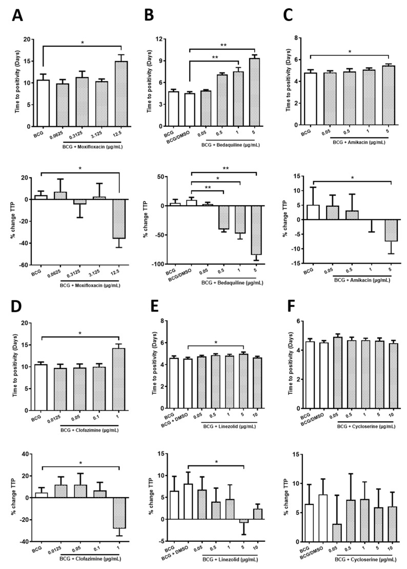
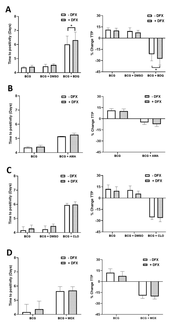
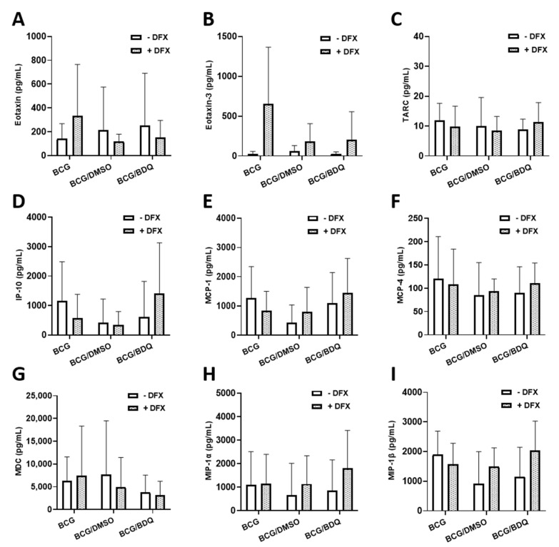
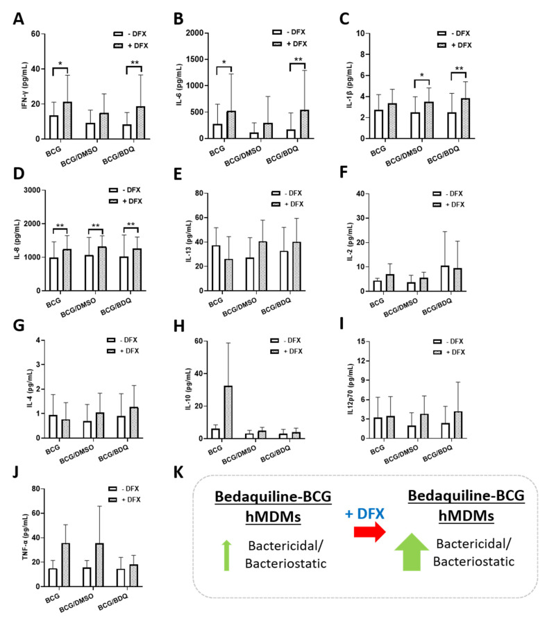
For over 50 years, patients with drug-sensitive and drug-resistant tuberculosis have undergone long, arduous, and complex treatment processes with several antimicrobials. With the prevalence of drug-resistant strains on the rise and new therapies for tuberculosis urgently required, we assessed whether manipulating iron levels in macrophages infected with mycobacteria offered some insight into improving current antimicrobials that are used to treat drug-resistant tuberculosis. We investigated if the iron chelator, desferrioxamine, can support the function of human macrophages treated with an array of second-line antimicrobials, including moxifloxacin, bedaquiline, amikacin, clofazimine, linezolid and cycloserine. Primary human monocyte-derived macrophages were infected with Bacillus Calmette-Guérin (BCG), which is pyrazinamide-resistant, and concomitantly treated for 5 days with desferrioxamine in combination with each one of the second-line tuberculosis antimicrobials. Our data indicate that desferrioxamine used as an adjunctive treatment to bedaquiline significantly reduced the bacterial load in human macrophages infected with BCG. Our findings also reveal a link between enhanced bactericidal activity and increases in specific cytokines, as the addition of desferrioxamine increased levels of IFN-γ, IL-6, and IL-1β in BCG-infected human monocyte-derived macrophages (hMDMs) treated with bedaquiline. These results provide insight, and an in vitro proof-of-concept, that iron chelators may prove an effective adjunctive therapy in combination with current tuberculosis antimicrobials.

Keywords: BCG; antimicrobials; drug-resistant tuberculosis; host-directed therapy; interferon-γ; iron chelation; iron metabolism; tuberculosis.


Conflict of interest statement

The authors declare no conflict of interest.

Figures

Figure 1
Figure 1
Determining the optimal concentration of moxifloxacin, bedaquiline, amikacin, clofazimine, linezolid and cycloserine in hMDMs infected with BCG. hMDMs, obtained from healthy blood donors, were infected with BCG for three hours and were subsequently treated with (A) moxifloxacin (0.0625, 0.3125, 3.125 and 12.5 µg/mL; n = 6), (B) bedaquiline (0.05, 0.5, 1 and 5 µg/mL; n = 3–4), (C) amikacin (0.05, 0.5, 1 or 5 µg/mL; n = 3–4), (D) clofazimine (0.0125, 0.05, 0.1 and 1 µg/mL; n = 4–5), (E) linezolid (0.05, 0.5, 1, 5 and 10 µg/mL; n = 3–7) or (F) cycloserine (0.05, 0.5, 1, 5 and 10 µg/mL; n = 3–7). Twenty-four hours post BCG infection, 25% of the cell supernatant was removed and the cells supplemented with 25% fresh cRPMI. Five days post infection, a mycobacterial growth inhibition assay (MGIA) was employed to investigate the capacity to control the growth of BCG in response to the second-line TB antimicrobials. Treatments deemed to increase time to positivity (TTP), shown in days, indicates a bactericidal or bacteriostatic effect; such inhibition in BCG growth can also be illustrated when plotted as percentage change in TTP (% change TTP; see methods section for description). The smallest antimicrobial concentration to significantly reduce TTP by >10% was chosen. Bars denote mean ± SEM. * p < 0.05 and ** p < 0.01 (mixed effects REML and Friedman ANOVA tests with Dunnett’s/Dunn’s multiple comparisons tests).
Figure 2
Figure 2
Examining if DFX can increase the efficacy of moxifloxacin, bedaquiline, amikacin and clofazimine in hMDMs infected with BCG. hMDMs, obtained from healthy blood donors, were infected with BCG for three hours and were subsequently treated with DFX (100 µM) in combination with (A) bedaquiline (0.5 µg/mL), (B) amikacin (5 µg/mL), (C) clofazimine (1 µg/mL) or (D) moxifloxacin (12.5 µg/mL). Twenty-four hours post BCG infection, 25% of the cell supernatant was removed, stored at −80 °C and the cells supplemented with 25% fresh cRPMI. Five days post infection, mycobacterial growth inhibition assays (MGIA) were undertaken to investigate the capacity to control the growth of BCG in response to the four antimicrobials. Treatments deemed to increase time to positivity (TTP), shown in days, indicates a bactericidal or bacteriostatic effect; such inhibition in BCG growth can also be illustrated when plotted as percentage change in TTP (% change TTP; see methods section for description). Bars denote mean± SEM. * p < 0.05 (Two-way repeated measures ANOVA tests with Šídák’s multiple comparisons tests, n = 5).
Figure 3
Figure 3
Assessing if dual DFX-bedaquiline treatment affects chemokine secretions in BCG-infected hMDMs. hMDMs, obtained from healthy blood donors, were infected with BCG for three hours and were subsequently treated with DFX (100 µM) in combination with bedaquiline (0.5 µg/mL). Twenty-four hours post BCG infection, cell supernatants were collected and levels of (A) eotaxin, (B) eotaxin-3, (C) TARC, (D) IP-10, (E) MCP-1, (F) MCP-4, (G) MDC, (H) MIP-1α and (I) MIP-1β were quantified using Meso Scale Discovery Multi-Array technology (n = 5). Bars denote mean ± SEM. p > 0.05 (Two-way repeated measures ANOVA tests with Šídák’s multiple comparisons tests).
Figure 4
Figure 4
Determining the effect of combined DFX-bedaquiline treatment on cytokine secretions in hMDMs infected with BCG. hMDMs, obtained from healthy blood donors, were infected with BCG for three hours and were subsequently treated with DFX (100 µM) in combination with bedaquiline (0.5 µg/mL). Twenty-four hours post BCG infection, cell supernatants were collected and levels of (A) IFNγ, (B) IL-6, (C) IL-1β, (D) IL-8, (E) IL-13, (F) IL-2, (G) IL-4, (H) IL-10, (I) IL12p70 and (J) TNF-α assayed using Meso Scale Discovery Multi-Array technology (n = 5). (K) DFX increases the bactericidal/bacteriostatic properties of bedaquiline in primary hMDMs infected with BCG. Bars denote mean ± SEM. * p < 0.05 and ** p < 0.01 (Two-way repeated measures ANOVA tests with Šídák’s multiple comparisons tests).

References

    1. Cahill C., Phelan J.J., Keane J. Understanding and Exploiting the Effect of Tuberculosis Antimicrobials on Host Mitochondrial Function and Bioenergetics. Front. Cell. Infect. Microbiol. 2020;10:493. doi: 10.3389/fcimb.2020.00493. - DOI - PMC - PubMed
    1. WHO . Global Tuberculosis Report 2020. World Health Organisation; Geneva, Switzerland: 2020.
    1. De Voss J.J., Rutter K.-L., Schroeder B.G., Su H., Zhu Y., Barry C.E. The salicylate-derived mycobactin siderophores of Mycobacterium tuberculosis are essential for growth in macrophages. Proc. Natl. Acad. Sci. USA. 2000;97:1252–1257. doi: 10.1073/pnas.97.3.1252. - DOI - PMC - PubMed
    1. Gomes M., Dom G., Pedrosa J., Boelaert J., Appelberg R. Effects of iron deprivation on Mycobacterium avium growth. Tuber. Lung Dis. 1999;79:321–328. doi: 10.1054/tuld.1999.0216. - DOI - PubMed
    1. Abreu R., Essler L., Giri P., Quinn F. Interferon-gamma promotes iron export in human macrophages to limit intracellular bacterial replication. PLoS ONE. 2020;15:e0240949. doi: 10.1371/journal.pone.0240949. - DOI - PMC - PubMed

MeSH terms

LinkOut - more resources