. 2021 Mar 25;26(7):1839.
doi: 10.3390/molecules26071839.

Chemokine Receptor 5 Antagonism Causes Reduction in Joint Inflammation in a Collagen-Induced Arthritis Mouse Model

Affiliations

Chemokine Receptor 5 Antagonism Causes Reduction in Joint Inflammation in a Collagen-Induced Arthritis Mouse Model

Mushtaq A Ansari et al. Molecules. .

Abstract

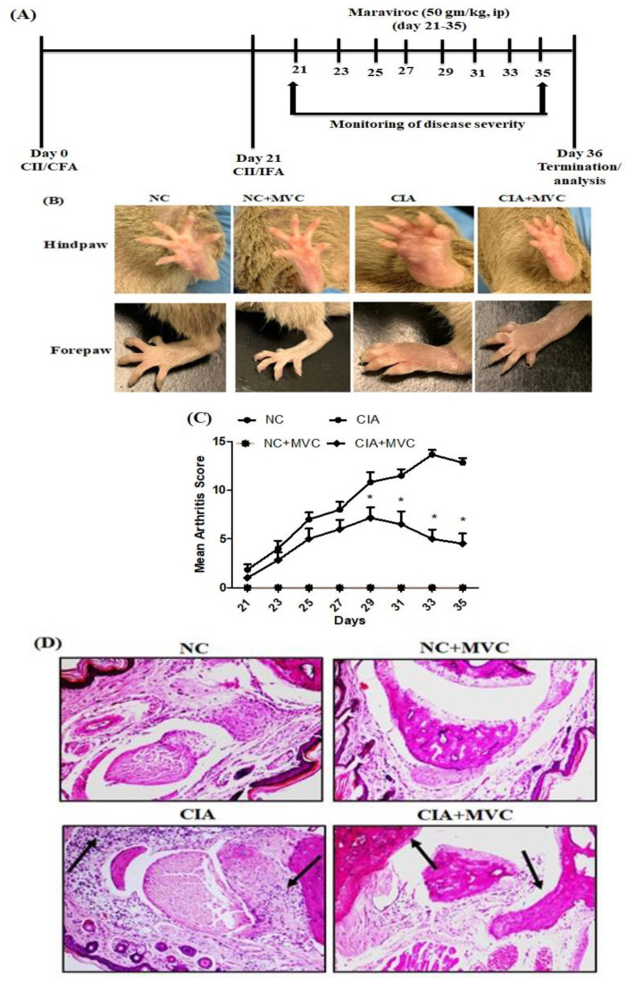
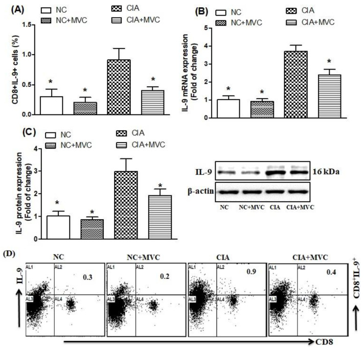
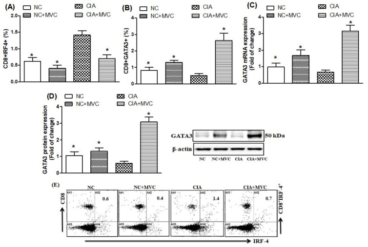
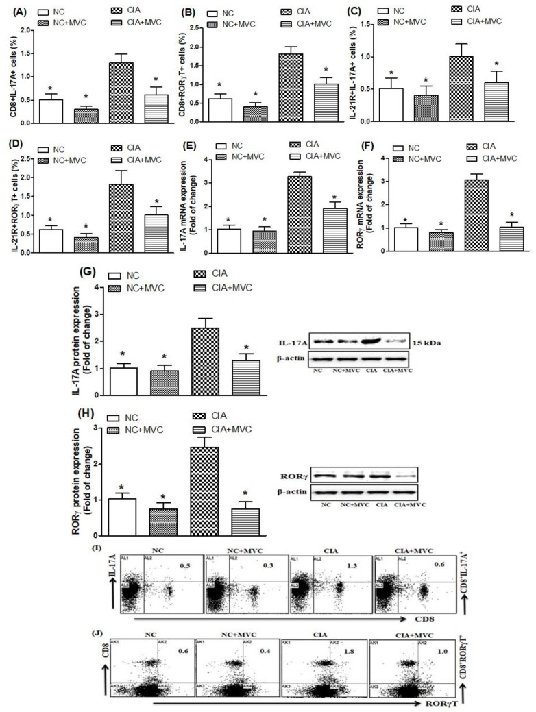
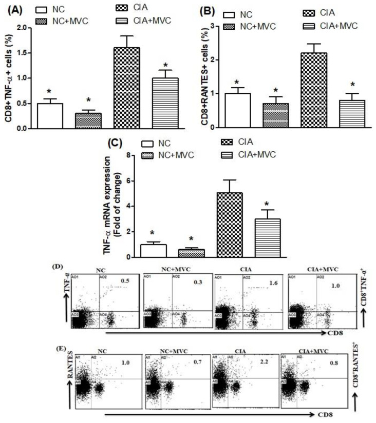
Rheumatoid arthritis (RA) is a chronic inflammatory disease mainly affecting the synovial joints. A highly potent antagonist of C-C chemokine receptor 5 (CCR5), maraviroc (MVC), plays an essential role in treating several infectious diseases but has not yet been evaluated for its potential effects on RA development. This study focused on evaluating the therapeutic potential of MVC on collagen-induced arthritis (CIA) in DBA/1J mice. Following CIA induction, animals were treated intraperitoneally with MVC (50 mg/kg) daily from day 21 until day 35 and evaluated for clinical score and histopathological changes in arthritic inflammation. We further investigated the effect of MVC on Th9 (IL-9, IRF-4, and GATA3) and Th17 (IL-21R, IL-17A, and RORγT) cells, TNF-α, and RANTES in CD8+ T cells in the spleen using flow cytometry. We also assessed the effect of MVC on mRNA and protein levels of IL-9, IL-17A, RORγT, and GATA3 in knee tissues using RT-PCR and western blot analysis. MVC treatment in CIA mice attenuated the clinical and histological severity of inflammatory arthritis, and it substantially decreased IL-9, IRF4, IL-21R, IL-17A, RORγT, TNF-α, and RANTES production but increased GATA3 production in CD8+ T cells. We further observed that MVC treatment decreased IL-9, IL-17A, and RORγt mRNA and protein levels and increased those of GATA3. This study elucidates the capacity of MVC to ameliorate the clinical and histological signs of CIA by reducing pro-inflammatory responses, suggesting that MVC may have novel therapeutic uses in the treatment of RA.

Keywords: CCR5 antagonist; CD8 cells; collagen-induced arthritis; inflammation; maraviroc.


Conflict of interest statement

The authors declare no conflict of interest.

Figures

Figure 1
Figure 1
MVC treatment prevents inflammation in collagen-induced arthritis (CIA) mice. DBA/1J mice were first immunized via intradermal injection of bovine type II collagen emulsified in complete Freund’s adjuvant. The second immunization was administered 21 days later in incomplete Freund’s adjuvant. (A) Work flow of MVC treatment for CIA in mice. Briefly, mice were first immunized with injection (s.c.) of emulsion containing bovine type II collagen (CII)/CFA on day 0 and were subsequently immunized with emulsion containing CII/IFA on day 21. Mice were treated with MVC from day 21 to day 35. The CIA mouse model was established, treated with MVC, and evaluated for clinical parameters of arthritis, including joint swelling (B), and mean arthritis score in CIA mice (C). Histological analysis of the joints showed a significant improvement in inflammation with less damage to the joint space in MVC-treated mice (D). Normal control (NC) mice received 1% (v/v) DMSO in saline and MVC (50 mg/kg) intraperitoneally (ip) daily from days 21 to 35. CIA mice were treated with MVC (50 mg/kg) ip after the second immunization. The level of significance was set at * p < 0.05 compared with the CIA control group. Data are presented as mean ± SD (n = 6).
Figure 2
Figure 2
(A) The effect of MVC on IL-9-producing CD8+ T cells was analyzed through flow cytometry in the spleen. (B,C) The expression levels of IL-9 mRNA (B) and protein (C) were analyzed by RT-PCR and western blot, respectively, in knee tissues from MVC-treated mice. (D) Representative dot plots of one mouse from each group. Normal control (NC) mice received 1% (v/v) DMSO in saline and MVC (50 mg/kg) intraperitoneally (ip) daily from days 21 to 35. CIA mice were treated with MVC (50 mg/kg) ip after the second immunization. The level of significance was set at * p < 0.05 compared with the CIA control group. Data are presented as mean ± SD (n = 6).
Figure 3
Figure 3
(A,B) The effects of MVC on IRF4- and GATA3-producing CD8+ T cells were analyzed through flow cytometry in the spleen. The expression levels of GATA3 mRNA (C) and protein (D) were analyzed by RT-PCR and western blot, respectively, in knee tissues from MVC-treated mice. (E) Representative dot plots of one mouse from each group. Normal control (NC) mice received 1% (v/v) DMSO in saline and MVC (50 mg/kg) intraperitoneally (ip) daily from days 21 to 35. CIA mice were treated with MVC (50 mg/kg) ip after the second immunization. The level of significance was set at * p < 0.05 compared with the CIA control group. Data are presented as mean ± SD (n = 6).
Figure 4
Figure 4
(AD). The effects of MVC on IL-17A- and RORγt-producing CD8+ T cells and IL-21+ receptor were analyzed through flow cytometry in the spleen. The mRNA expression levels of IL-17A (E) and RORγt (F) were analyzed by RT-PCR in knee tissues from MVC-treated mice. The protein expression levels of IL-17A (G) and RORγt (H) were analyzed by western blot in knee tissues from MVC-treated mice. (I,J) Representative dot plots of one mouse from each group. Normal control (NC) mice received 1% (v/v) DMSO in saline and MVC (50 mg/kg) intraperitoneally (ip) daily from days 21 to 35. CIA mice were treated with MVC (50 mg/kg) ip after the second immunization. The level of significance was set at * p < 0.05 compared with the CIA control group. Data are presented as mean ± SD (n = 6).
Figure 5
Figure 5
(A,B). The effects of MVC on TNF-α- and RANTES-producing CD8+ T cells were analyzed through flow cytometry in the spleen. (C) The mRNA expression levels of TNF-α was analyzed by RT-PCR in knee tissues from MVC-treated mice. (D,E) Representative dot plots of one mouse from each group. Normal control (NC) mice received 1% (v/v) DMSO in saline and MVC (50 mg/kg) intraperitoneally (ip) daily from days 21 to 35. CIA mice were treated with MVC (50 mg/kg) ip after the second immunization. The level of significance was set at * p < 0.05 compared with the CIA control group. Data are presented as mean ± SD (n = 6).

Similar articles

Cited by

References

    1. Iwamoto T., Okamoto H., Toyama Y., Momohara S. Molecular aspects of rheumatoid arthritis: Chemokines in the joints of pa-tients. FEBS J. 2008;275:4448–4455. doi: 10.1111/j.1742-4658.2008.06580.x. - DOI - PubMed
    1. Firestein G.S. Evolving concepts of rheumatoid arthritis. Nat. Cell Biol. 2003;423:356–361. doi: 10.1038/nature01661. - DOI - PubMed
    1. Scott D.L., Wolfe F., Huizinga T.W. Rheumatoid arthritis. Lancet. 2010;376:1094–1108. doi: 10.1016/S0140-6736(10)60826-4. - DOI - PubMed
    1. Park J.S., Moon S.J., Lim M., Byun J.K., Hwang S.H., Yang S., Kim E.K., Kim S.-M., Lee J., Kwok S., et al. Retinoic Acid Receptor-Related Receptor Alpha Ameliorates Autoimmune Arthritis via Inhibiting of Th17 Cells and Osteoclastogenesis. Front. Immunol. 2019;10:2270. doi: 10.3389/fimmu.2019.02270. - DOI - PMC - PubMed
    1. Angelotti F., Parma A., Cafaro G., Capecchi R., Alunno A., Puxeddu I. One year in review 2017: Pathogenesis of rheumatoid arthritis. Clin. Exp. Rheumatol. 2017;35:368–378. - PubMed

MeSH terms

LinkOut - more resources