Review
. 2021 Mar 25;22(7):3366.
doi: 10.3390/ijms22073366.

Exogenous Integrin αIIbβ3 Inhibitors Revisited: Past, Present and Future Applications

Affiliations
Review

Exogenous Integrin αIIbβ3 Inhibitors Revisited: Past, Present and Future Applications

Danique L van den Kerkhof et al. Int J Mol Sci. .

Abstract

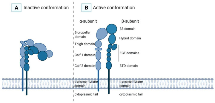
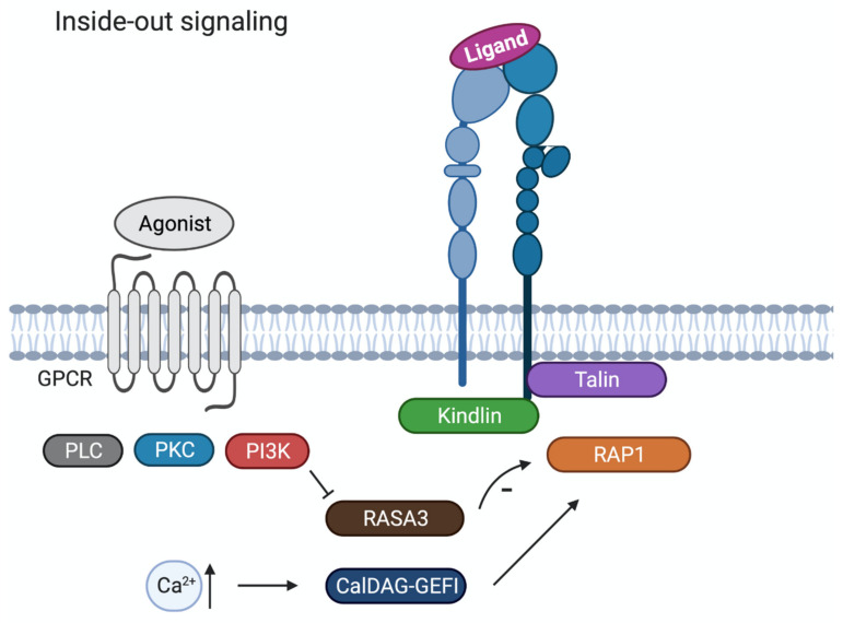
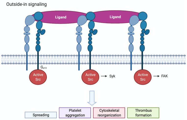
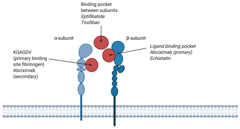
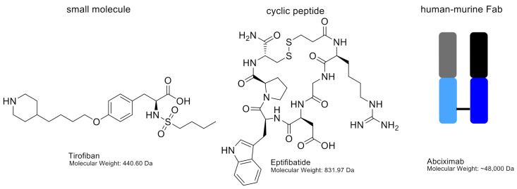
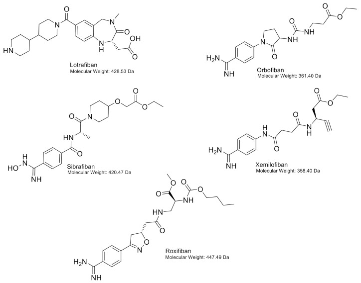
The integrin αIIbβ3 is the most abundant integrin on platelets. Upon platelet activation, the integrin changes its conformation (inside-out signalling) and outside-in signalling takes place leading to platelet spreading, platelet aggregation and thrombus formation. Bloodsucking parasites such as mosquitoes, leeches and ticks express anticoagulant and antiplatelet proteins, which represent major sources of lead compounds for the development of useful therapeutic agents for the treatment of haemostatic disorders or cardiovascular diseases. In addition to hematophagous parasites, snakes also possess anticoagulant and antiplatelet proteins in their salivary glands. Two snake venom proteins have been developed into two antiplatelet drugs that are currently used in the clinic. The group of proteins discussed in this review are disintegrins, low molecular weight integrin-binding cysteine-rich proteins, found in snakes, ticks, leeches, worms and horseflies. Finally, we highlight various oral antagonists, which have been tested in clinical trials but were discontinued due to an increase in mortality. No new αIIbβ3 inhibitors are developed since the approval of current platelet antagonists, and structure-function analysis of exogenous disintegrins could help find platelet antagonists with fewer adverse side effects.

Keywords: antagonists; hematophagous parasites; integrin αIIbβ3; platelets.


Conflict of interest statement

The authors declare no conflict of interest.

Figures

Figure 1
Figure 1
In the inactive conformation of the αIIbβ3 integrin (A), the ligand-binding site is poorly accessible for its ligands, while in the active conformation (B) the ligand-binding site is exhibited and has high affinity for its ligands.
Figure 2
Figure 2
Schematic overview of inside-out signalling in platelets. Agonist activation of G protein-coupled receptor triggers signalling pathways with key signalling proteins like phospholipase C (PLC), protein kinase C (PKC) and phosphatidylinositide-3-kinase (PI3K). Increased Ca2+ will lead to activation of CalDAG-GEFI that will activate Ras-related protein 1 (RAP1). RASA3 acts as a negative regulator of RAP1 activation, and RASA3 activity is restricted by PI3K activation. Through the separation of the talin head and tail domain, the talin head domain associates with the cytoplasmic tail of the β3 subunit, converting the integrin αIIbβ3 from the inactive to the active form.
Figure 3
Figure 3
Schematic overview of outside-in signalling in platelets. Clustered integrins will initiate outside-in signalling. c-Src associates with the β3-integrin tail and becomes activated. Src, Syk and FAK will regulate downstream signalling via tyrosine phosphorylation. Outside-in signalling leads to spreading, platelet aggregation, cytoskeletal reorganisation and thrombus formation.
Figure 4
Figure 4
Different binding sites of antagonists on αIIbβ3 integrins. KQAGDV; the primary binding site for fibrinogen and a secondary binding site for abciximab. The binding pocket between subunits; binding site for eptifibatide and tirofiban, therefore competing with fibrinogen. Binding pocket β3; primary binding site for abciximab and echistatin, therefore not competing with fibrinogen.
Figure 5
Figure 5
Chemical structures of tirofiban and eptifibatide, including molecular weights. Abciximab is shown as a schematic figure. The murine variable chain region is shown in grey (light chain) and black (heavy chain). The human constant regions are shown in light blue (light chain) and dark blue (heavy chain).
Figure 6
Figure 6
Chemical structures of lotrafiban, orbofiban, sibrafiban, xemilofiban and roxifiban, including molecular weights.

References

    1. Huang J., Li X., Shi X., Zhu M., Wang J., Huang S., Huang X., Wang H., Li L., Deng H., et al. Platelet integrin alphaIIbbeta3: Signal transduction, regulation, and its therapeutic targeting. J. Hematol. Oncol. 2019;12:26. doi: 10.1186/s13045-019-0709-6. - DOI - PMC - PubMed
    1. Anderson L.R., Owens T.W., Naylor M.J. Structural and mechanical functions of integrins. Biophys. Rev. 2014;6:203–213. doi: 10.1007/s12551-013-0124-0. - DOI - PMC - PubMed
    1. Zhang K., Chen J. The regulation of integrin function by divalent cations. Cell Adhes. Migr. 2012;6:20–29. doi: 10.4161/cam.18702. - DOI - PMC - PubMed
    1. Bazzoni G., Shih D.T., Buck C.A., Hemler M.E. Monoclonal antibody 9EG7 defines a novel beta 1 integrin epitope induced by soluble ligand and manganese, but inhibited by calcium. J. Biol. Chem. 1995;270:25570–25577. doi: 10.1074/jbc.270.43.25570. - DOI - PubMed
    1. Heino J. The collagen family members as cell adhesion proteins. Bioessays. 2007;29:1001–1010. doi: 10.1002/bies.20636. - DOI - PubMed

MeSH terms