. 2021 Mar 25;22(7):3386.
doi: 10.3390/ijms22073386.

Ligand-Induced GPR110 Activation Facilitates Axon Growth after Injury

Affiliations

Ligand-Induced GPR110 Activation Facilitates Axon Growth after Injury

Heungsun Kwon et al. Int J Mol Sci. .

Abstract

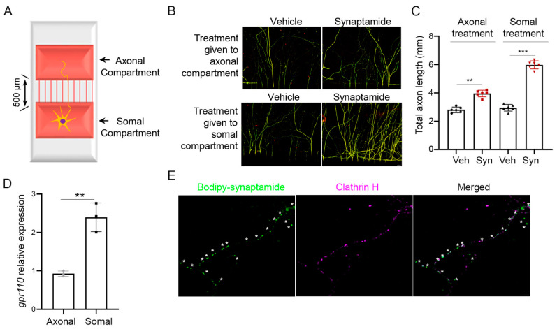
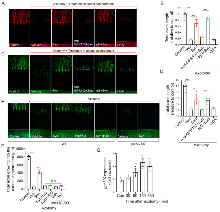
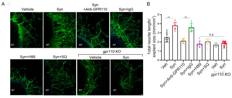
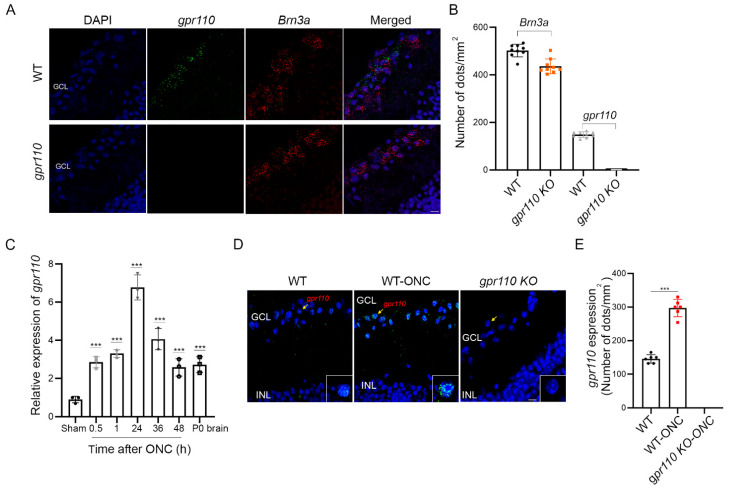
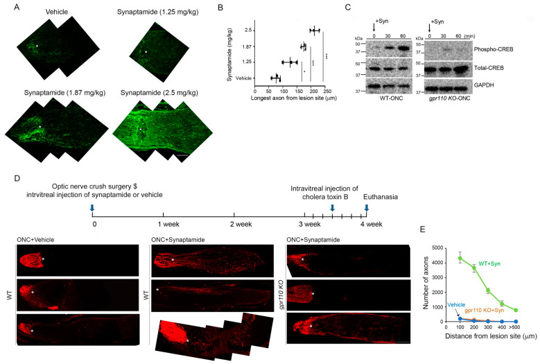
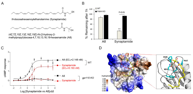
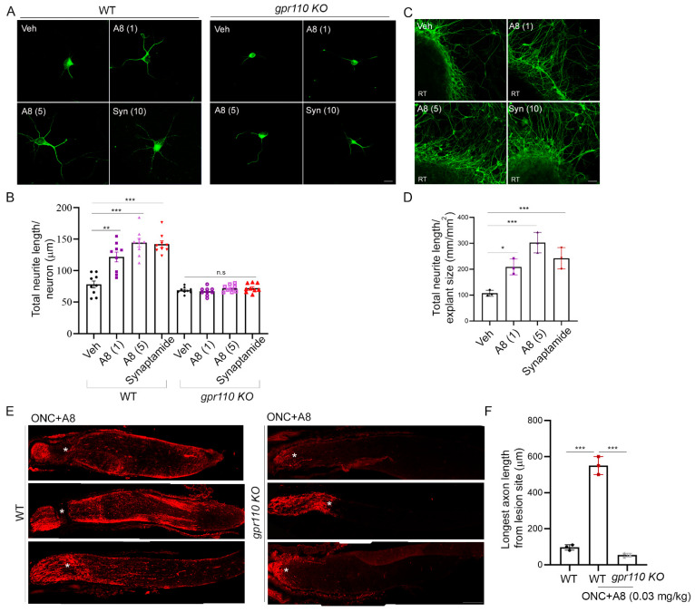
Recovery from axonal injury is extremely difficult, especially for adult neurons. Here, we demonstrate that the activation of G-protein coupled receptor 110 (GPR110, ADGRF1) is a mechanism to stimulate axon growth after injury. N-docosahexaenoylethanolamine (synaptamide), an endogenous ligand of GPR110 that promotes neurite outgrowth and synaptogenesis in developing neurons, and a synthetic GPR110 ligand stimulated neurite growth in axotomized cortical neurons and in retinal explant cultures. Intravitreal injection of GPR110 ligands following optic nerve crush injury promoted axon extension in adult wild-type, but not in gpr110 knockout, mice. In vitro axotomy or in vivo optic nerve injury rapidly induced the neuronal expression of gpr110. Activating the developmental mechanism of neurite outgrowth by specifically targeting GPR110 that is upregulated upon injury may provide a novel strategy for stimulating axon growth after nerve injury in adults.

Keywords: GPR110; axon; cAMP/PKA; microfluidic culture platform; optic nerve; retinal explant culture; synaptamide.


Conflict of interest statement

The authors declare no conflict of interest.

Figures

Figure 1
Figure 1
Axon growth promoted by either somal or axonal treatment of synaptamide. (A) Schematic representation of a microfluidic culture platform with two chambers separated by 500 μm of thin grooves that was used to culture primary cortical neurons from the mouse brain at postnatal day 0 (P0). (B) β-III tubulin immunofluorescence micrographs showing that axon outgrowth in primary cortical neurons was induced by the application of 10 nM synaptamide to the axonal or somal compartment on DIV3 and cultured for an additional 4 days. (C) Quantitative results of the total length of axons entered in the axonal compartment. Synaptamide application to the cell body or axonal compartment resulted in a 2.2- or 1.3-fold increase in axon growth, respectively, compared to the vehicle control. (D) Relative quantification of gpr110 expression in soma and axon determined by RT-PCR after collecting samples separately from the somal or axonal compartment of the two-chamber culture platform. (E) Internalization of GPR110 visualized by a fluorescent ligand bodipy–synaptamide. After 30 min stimulation of DIV 3 cortical neurons with 20 nM bodipy-synaptamide, internalized GPR110 was detected as fluorescent endocytic puncta (green) overlapping with an endocytic marker clathrin H (magenta), confirming the axonal presence of GPR110. Asterisks (*) represent endocytic GPR110 receptor puncta. Data are expressed as mean ± s.e.m. with n = 6 for (B) and n = 3 for (D) per group. ** p < 0.01, *** p < 0.001 by t-test; Veh, Vehicle (DMSO); Syn, synaptamide. Scale bar, 20 μm (B), 1 μm (E).
Figure 2
Figure 2
Synaptamide stimulates axon regeneration in vitro through GPR110/cAMP/PKA-dependent signaling. (AD), Stimulated axon regrowth by somal (A,B) or axonal treatment (C,D) with 10 nM synaptamide, visualized by β-III tubulin immunostaining and quantified by measuring the total axon length in the axonal compartment 7 days after axotomy. (E,F), Effect of cAMP (SQ22536, 10 µM) and PKA (H89, 10 µM) inhibitors, N-terminal targeting GPR110 antibody (0.4 mg/mL), or gpr110 KO on synaptamide-induced axon regrowth with quantitative results normalized to the non-axotomized control. G, Time-dependence of axotomy-induced increase in gpr110 expression measured by qRT-PCR. Data are expressed as mean ± s.e.m. with n = 9 for (B,D,F) and n = 6 for (G) per group. ** p < 0.01, *** p < 0.001, Con, non-axotomized control; Veh, vehicle (DMSO); Syn, synaptamide. Scale bars, 20 μm. n.s, statistically not significant.
Figure 3
Figure 3
Retinal neurite outgrowth is stimulated by synaptamide through the GPR110/cAMP/PKA signaling pathway. (A), Neurite outgrowth from cultured P0 retinal explants increased by 10 nM synaptamide and inhibited by N-terminal targeting GPR110 antibody (0.4 mg/mL), SQ22536 (10 µM), H89 (10 µM), or gpr110 KO. The cultures were treated with agents on DIV 3 for 7 days and immunostained for β-III tubulin. (B), Total neurite length per field adjusted for the explant size. Data are expressed as mean ± s.e.m with n = 9 per group. ** p < 0.01 by one-way ANOVA. RT, retinal explant tissue. Scale bar, 50 μm. n.s, statistically not significant.
Figure 4
Figure 4
Optic nerve injury upregulates gpr110 expression. (A) Expression of gpr110 in RGCs from 4-month-old mice detected by in situ hybridization using probes for gpr110 (green) and a RGC marker Brn3a (red), overlaid on DAPI-stained nuclei (blue). (B) Quantitation of gpr110 expression. (CE) Induction of gpr110 in the retina tissue after ONC determined by qRT-PCR (C) and by in situ hybridization of the ganglion cell layer (GCL) with micrographic (D) and quantitative results (E). Data are expressed as mean ± s.e.m with n = 9 (B) n = 3 (C) and n = 6 (E) per group. *** p < 0.001 by one-way ANOVA. GCL, ganglion cell layer; INL, inner nuclear layer. Scale bars, 20 μm (A,D).
Figure 5
Figure 5
Synaptamide GPR110-dependently induces axon growth after optic nerve injury. (A) Dose-dependent effect of synaptamide on stimulated axon growth after injury. ONC was performed on 4-month-old BALB/C mice and synaptamide was administrated intravitreally at the doses indicated. Longitudinal sections through the optic nerve were collected at 4 weeks after ONC and regenerating axons were visualized by anti-GAP43 antibody (green, 1:500). Lesion site was marked by asterisks (*). (B) Quantitative measurement of axon length extended from the lesion site after synaptamide injection at the doses indicated. (C) Time-dependent increase in CREB phosphorylation after synaptamide injection into 4-month-old WT but not gpr110 KO mouse retinas following ONC. (D) Optic nerve tissue sections collected at 4 weeks after ONC and visualized by CTB, indicating that intravitreal injection of synaptamide (2.5 mg/kg) induced axon growth in WT but not in gpr110 KO mice. The optic nerve micrographs shown in A and at the bottom panel of the ONC+Synaptamide group in D were obtained after the manual stitching of image sections using Photoshop. (E) The number of the axons extended to a 100–600 micrometer distance from the lesion site, indicated by the asterisk. Data are expressed as mean ± s.e.m with n = 3 (A) and n = 4 (E) per group. * p < 0.05, *** p < 0.001 by one-way ANOVA. Scale bars, 100 μm (A,D).
Figure 6
Figure 6
A8, a FAAH-resistant structural analogue of synaptamide, exhibited improved cAMP production and receptor binding. (A) Chemical structures of synaptamide and its stable analogue A8 ((4Z,7Z,10Z,13Z,16Z,19Z)-N-(2-hydroxy-2-methylpropyl)docosa-4,7,10,13,16,19-hexaenamide). (B) Synaptamide and A8 remaining after incubation with brain homogenates from WT or fatty acid amide hydrolase (FAAH) KO mice for 1 h, showing that A8 is resistant to FAAH hydrolysis. (C) Dose-dependent increase in cAMP production elicited by synaptamide and A8. Cortical neurons derived from P0 brains were stimulated for 15 min on DIV3 for cAMP measurement. (D) Improved binding ability of A8 to GPR110 predicted by the binding model of A8 at the GAIN domain of GPR110. The protein surface is rendered in the color of hydrophobicity. A8 is shown in sticks of yellow carbon atoms, and key residues at the hydrophobic pocket are shown in sticks of cyan carbon atoms. O and N atoms are shown in red and blue, respectively. The two methyl groups (labeled with 1 and 2) of A8 fit well in the hydrophobic pocket formed by P470 and P417. The resulting hydrophobic interaction stabilizes the hydrogen bonding between the polar moieties of GPR110 and synaptamide or A8 (dotted yellow lines). ** p < 0.01 vs. A8.
Figure 7
Figure 7
A8, a FAAH-resistant structural analogue of synaptamide, improved neurite outgrowth and axon growth after injury. (A,B) Improved neurite outgrowth by A8 in cultured cortical neurons from WT but not from gpr110 KO mice, indicating the GPR110-dependent nature of A8 bioactivity. Neurite length was evaluated after treating DIV0 cultures with synaptamide (10 nM) or A8 (1 or 5 nM) for 3 days. (C,D) RGC neurite growth in retinal explant cultures promoted by synaptamide or A8 as indicated by the quantification of axon length after immunostaining with anti β-III tubulin antibody. (E,F) GPR110-dependent stimulatory effect of intravitreal injection of A8 at 0.03 mg/kg on axon extension, shown by the micrographic images of CTB-labeled axons (E) and quantitative data showing the longest axon length (F) obtained at 4 weeks after ONC. Some of the optic nerve micrographs (E) were obtained after the manual stitching of image sections using Photoshop. Data are expressed as mean ± s.e.m with n = 9 (A) and n = 3 (D,F) per group. RT, retinal tissue; * p < 0.05, ** p < 0.01, *** p < 0.001; Scale bars, 10 μm (A), 50 μm (C) and 100 μm (E).

References

    1. Fischer D., Leibinger M. Promoting optic nerve regeneration. Prog. Retin. Eye Res. 2012;31:688–701. doi: 10.1016/j.preteyeres.2012.06.005. - DOI - PubMed
    1. He Z., Jin Y. Intrinsic Control of Axon Regeneration. Neuron. 2016;90:437–451. doi: 10.1016/j.neuron.2016.04.022. - DOI - PubMed
    1. Silver S.J., Schwab M.E., Popovich P.G. Central Nervous System Regenerative Failure: Role of Oligodendrocytes, Astrocytes, and Microglia. Cold Spring Harb. Perspect. Biol. 2015;7:a020602. doi: 10.1101/cshperspect.a020602. - DOI - PMC - PubMed
    1. Berkelaar M., Clarke D.B., Wang Y.C., Bray G.M., Aguayo A.J. Axotomy results in delayed death and apoptosis of retinal ganglion cells in adult rats. J. Neurosci. 1994;14:4368–4374. doi: 10.1523/JNEUROSCI.14-07-04368.1994. - DOI - PMC - PubMed
    1. McGee A.W., Yang Y., Fischer Q.S., Daw N.W., Strittmatter S.M. Experience-driven plasticity of visual cortex limited by myelin and Nogo receptor. Science. 2005;309:2222–2226. doi: 10.1126/science.1114362. - DOI - PMC - PubMed

LinkOut - more resources