. 2021 Mar 4;13(5):1090.
doi: 10.3390/cancers13051090.

Pancreatic Cancers with High Grade Tumor Budding Exhibit Hallmarks of Diminished Anti-Tumor Immunity

Affiliations

Pancreatic Cancers with High Grade Tumor Budding Exhibit Hallmarks of Diminished Anti-Tumor Immunity

Hassan Sadozai et al. Cancers (Basel). .

Abstract

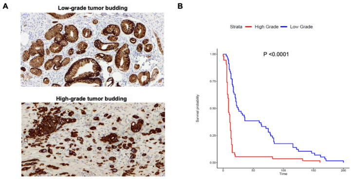
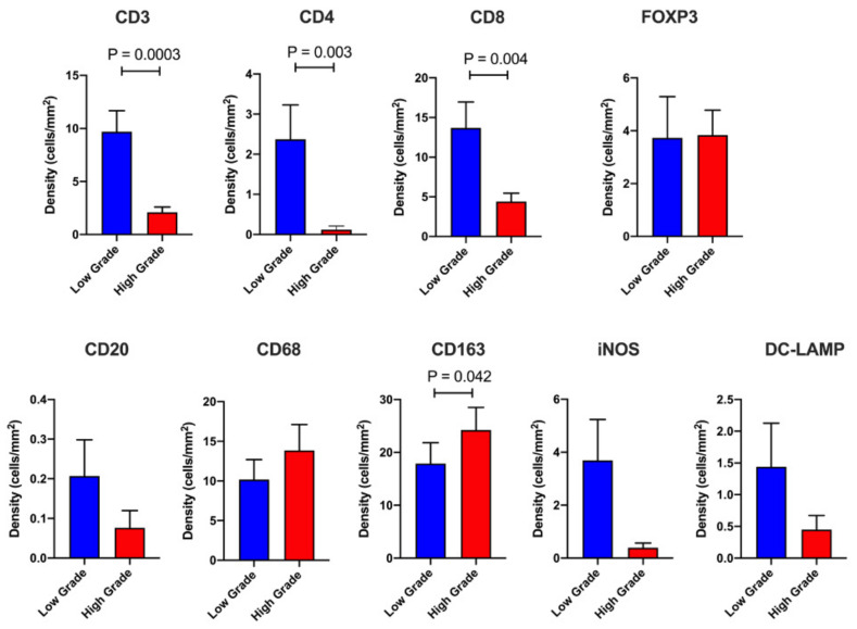
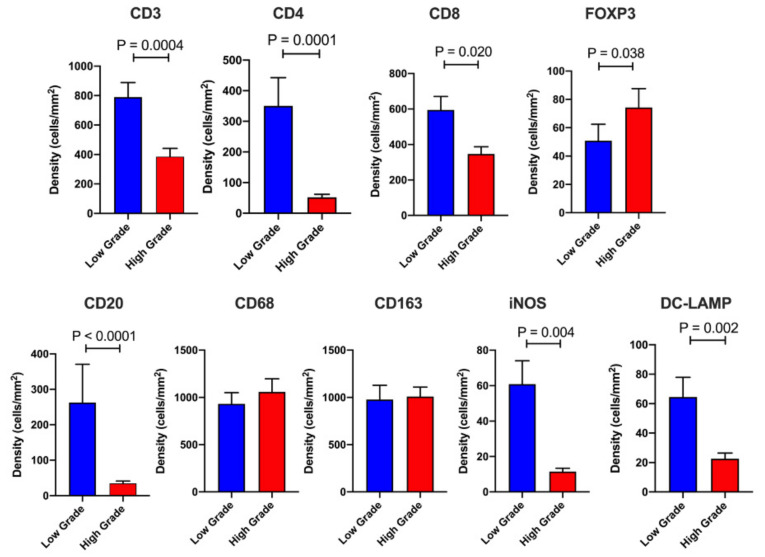
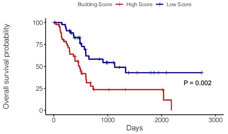
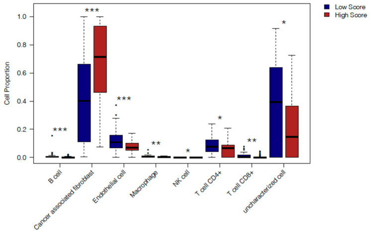
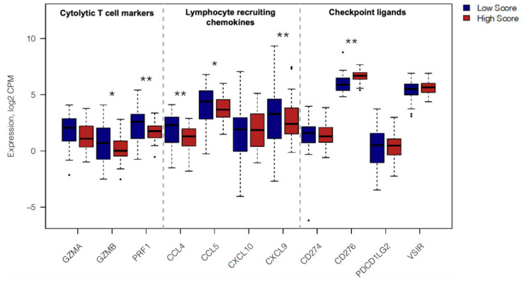
Tumor budding is associated with epithelial-mesenchymal transition and diminished survival in a number of cancer types including pancreatic ductal adenocarcinoma (PDAC). In this study, we dissect the immune landscapes of patients with high grade versus low grade tumor budding to determine the features associated with immune escape and disease progression in pancreatic cancer. We performed immunohistochemistry-based quantification of tumor-infiltrating leukocytes and tumor bud assessment in a cohort of n = 111 PDAC patients in a tissue microarray (TMA) format. Patients were divided based on the ITBCC categories of tumor budding as Low Grade (LG: categories 1 and 2) and High Grade (HG: category 3). Tumor budding numbers and tumor budding grade demonstrated a significant association with diminished overall survival (OS). HG cases exhibit notably reduced densities of stromal (S) and intratumoral (IT) T cells. HG cases also display lower M1 macrophages (S) and increased M2 macrophages (IT). These findings were validated using gene expression data from TCGA. A published tumor budding gene signature demonstrated a significant association with diminished survival in PDAC patients in TCGA. Immune-related gene expression revealed an immunosuppressive TME in PDAC cases with high expression of the budding signature. Our findings highlight a number of immune features that permit an improved understanding of disease progression and EMT in pancreatic cancer.

Keywords: M1/M2 macrophages; T cell-enriched; T lymphocytes; gene signature; pancreatic ductal adenocarcinoma (PDAC); tumor budding.


Conflict of interest statement

The authors declare no conflict of interest.

Figures

Figure 1
Figure 1
Tumor budding grade is associated with overall survival in PDAC. (A) Representative images of cases from High Grade vs. Low Grade tumor budding groups. (B) Kaplan-Meier curves comparing the OS of PDAC cases with High Grade (Category 3) versus Low Grade (Category 1&2) tumor budding. There were n = 54 cases in the High Grade versus n = 57 cases in the Low Grade budding group. Statistical comparisons were performed using the log-rank test.
Figure 2
Figure 2
Intratumoral (IT) immune profiling. Barplots displaying the IT leukocyte density in High Grade (n = 54) versus Low Grade (n = 57) tumor budding cases. The following immune cell markers were utilized (CD3, CD4, CD8, FOXP3, CD20, CD68, iNOS, CD163, DC-LAMP). Data are depicted as mean + standard error (SE). Differences between groups were analyzed using the Mann-Whitney U test. Only significant p-values (<0.05) are shown on the graph.
Figure 3
Figure 3
Stromal (S) immune profiling. Barplots displaying the S leukocyte density in High Grade (n = 54) versus Low Grade (n = 57) tumor budding cases. The following immune cell markers were utilized (CD3, CD4, CD8, FOXP3, CD20, CD68, iNOS, CD163, DC-LAMP). Data are depicted as mean + SE. Differences between groups were analyzed using the Mann-Whitney U test. Only significant P values are shown on the graph.
Figure 4
Figure 4
Prognostic role of tumor budding gene signature. Kaplan-Meier curves comparing the OS of PDAC cases from TCGA with a High (red) vs. Low (blue) Score for the molecular budding signature. Cases were dichotomized as high or low by top and bottom quartiles of expression (n = 45 patients each). Statistical comparisons were performed using the log-rank test. The budding gene score was computed using the singscore package in Bioconductor (see methods).
Figure 5
Figure 5
Cell type deconvolution of TCGA PDAC cases with High and Low Budding gene signature scores. Boxplots comparing the cell fractions as computed by the EPIC algorithm for immune and non-immune cells (B cells, CAFs, Endothelial cells, Macrophages, NK cells, CD4+ T cells, CD8+ T cells and uncharacterized cells) in High vs. Low Budding score cases (n = 45 patients each) from TCGA. Data are shown as box and whisker plots with the box representing the IQR and the whiskers extending to the minimum and maximum values. Statistical comparisons between High and Low Score cases were performed for each immune cell subset individually using the Mann-Whitney U test. Only significant p values (<0.05) are shown on the graph. *** p < 0.001, ** p < 0.01, * p < 0.05.
Figure 6
Figure 6
Immune related gene expression in TCGA PDAC cases with High and Low Budding gene signature scores. Boxplots comparing gene expression of immunological genes in High and Low Budding Score patients from TCGA (n = 45 patients each). Gene expression is shown as log2-transformed, normalized CPM (counts per million) values. Transcriptome-wide differential expression statistics were computed using EdgeR under quasi-likelihood framework. P values were corrected for multiple testing using the Benjamini-Hochberg method limiting the correction to the 11 genes of interest displayed above. Only significant p values (<0.05) are shown on the graph. ** p < 0.01, * p < 0.05.

References

    1. Kamisawa T., Wood L.D., Itoi T., Takaori K. Pancreatic cancer. Lancet. 2016;388:73–85. doi: 10.1016/S0140-6736(16)00141-0. - DOI - PubMed
    1. Strobel O., Neoptolemos J., Jäger D., Büchler M.W. Optimizing the outcomes of pancreatic cancer surgery. Nat. Rev. Clin. Oncol. 2019;16:11–26. doi: 10.1038/s41571-018-0112-1. - DOI - PubMed
    1. Collisson E.A., Sadanandam A., Olson P., Gibb W.J., Truitt M., Gu S., Cooc J., Weinkle J., Kim G.E., Jakkula L., et al. Subtypes of pancreatic ductal adenocarcinoma and their differing responses to therapy. Nat. Med. 2011;17 doi: 10.1038/nm.2344. - DOI - PMC - PubMed
    1. Raut C.P., Tseng J.F., Sun C.C., Wang H., Wolff R.A., Crane C.H., Hwang R., Vauthey J.-N., Abdalla E.K., Lee J.E., et al. Impact of Resection Status on Pattern of Failure and Survival After Pancreaticoduodenectomy for Pancreatic Adenocarcinoma. Ann. Surg. 2007;246:52–60. doi: 10.1097/01.sla.0000259391.84304.2b. - DOI - PMC - PubMed
    1. Sierzega M., Popiela T., Kulig J., Nowak K. The Ratio of Metastatic/Resected Lymph Nodes is an Independent Prognostic Factor in Patients with Node-positive Pancreatic Head Cancer. Pancreas. 2006;33:240–245. doi: 10.1097/01.mpa.0000235306.96486.2a. - DOI - PubMed