. 2021 Mar 4;26(5):1362.
doi: 10.3390/molecules26051362.

Fluorine-19 Magnetic Resonance Imaging for Detection of Amyloid β Oligomers Using a Keto Form of Curcumin Derivative in a Mouse Model of Alzheimer's Disease

Affiliations

Fluorine-19 Magnetic Resonance Imaging for Detection of Amyloid β Oligomers Using a Keto Form of Curcumin Derivative in a Mouse Model of Alzheimer's Disease

Daijiro Yanagisawa et al. Molecules. .

Abstract

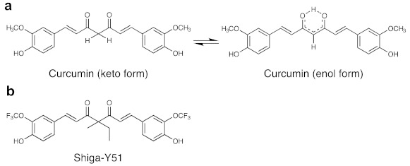
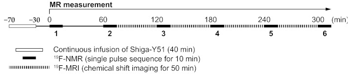
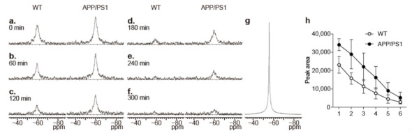
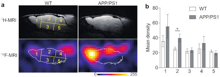
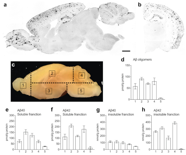
Recent evidence suggests that the formation of soluble amyloid β (Aβ) aggregates with high toxicity, such as oligomers and protofibrils, is a key event that causes Alzheimer's disease (AD). However, understanding the pathophysiological role of such soluble Aβ aggregates in the brain in vivo could be difficult due to the lack of a clinically available method to detect, visualize, and quantify soluble Aβ aggregates in the brain. We had synthesized a novel fluorinated curcumin derivative with a fixed keto form, named as Shiga-Y51, which exhibited high selectivity to Aβ oligomers in vitro. In this study, we investigated the in vivo detection of Aβ oligomers by fluorine-19 (19F) magnetic resonance imaging (MRI) using Shiga-Y51 in an APP/PS1 double transgenic mouse model of AD. Significantly high levels of 19F signals were detected in the upper forebrain region of APP/PS1 mice compared with wild-type mice. Moreover, the highest levels of Aβ oligomers were detected in the upper forebrain region of APP/PS1 mice in enzyme-linked immunosorbent assay. These findings suggested that 19F-MRI using Shiga-Y51 detected Aβ oligomers in the in vivo brain. Therefore, 19F-MRI using Shiga-Y51 with a 7 T MR scanner could be a powerful tool for imaging Aβ oligomers in the brain.

Keywords: Alzheimer’s disease; curcumin; imaging biomarker; keto-enol tautomerism; magnetic resonance imaging; mouse model.


Conflict of interest statement

The authors declare no conflict of interest.

Figures

Figure 1
Figure 1
Chemical structures of curcumin and Shiga-Y51. (a) Curcumin, a yellow-orange pigment in turmeric, exists in an equilibrium between keto and enol tautomers. (b) Shiga-Y51 is a fluorinated curcumin derivative with a fixed keto form by the methyl and ethyl groups at the C4 position.
Figure 2
Figure 2
Timeline of the present study. Shiga-Y51 (200 mg/kg, i.v.) was injected by a continuous infusion over 40 min in APP/PS1 and wild-type mice under deep anesthesia. The first fluorine-19 nuclear magnetic resonance (19F-NMR) measurement was performed 30 min after the injection and repeated every 60 min. Fluorine-19 chemical shift imaging (19F-CSI) data for the fluorine-19 magnetic resonance (19F-MR) images were collected for 50 min every 1 h after the second 19F-NMR measurement.
Figure 3
Figure 3
19F-NMR spectra from the mouse head. (af) Representative 19F-NMR spectra from the mouse heads of wild-type (WT) and APP/PS1 mice that were obtained every 60 min from 30 min after the injection. The spectra in (a) to (f) were from the measurements 1 to 6 in Figure 2, respectively. (g) 19F-NMR spectra of Shiga-Y51 dissolved in DMSO. Chemical shifts (ppm) were referenced to C6F6 at −163 ppm as an external standard. The FID signal was processed with 40 Hz line broadening similarly to CSI data. The line width of 19F signal in the 19F-NMR of Shiga-Y51 dissolved in DMSO was 177 Hz, whereas the 19F-NMR spectra from the mouse head showed the line width of 760 Hz. (h) The time course of changes in the peak area was measured in WT (n = 4) and APP/PS1 mice (n = 3). Data are presented as mean ± standard error of mean (SEM).
Figure 4
Figure 4
Accumulation of 19F signals in the brain of APP/PS1 mice. (a) Representative 19F-MR images in wild-type (WT) and APP/PS1 mice that were obtained for 50 min in the first 19F-CSI measurement (at 100 min after the injection). A lookup table (LUT) was used to display the 19F-MRI signal, and the white lines in each 19F-MR image indicate the outline of the brain delineated based on the corresponding 1H-MR images. (b) The levels of 19F signals were measured in five regions indicated in yellow color. Significantly high levels of 19F signals were detected in region 2 in APP/PS1 mice (n = 3) compared with WT mice (n = 4). Data are presented as mean ± SEM. * p < 0.05 (unpaired t-test).
Figure 5
Figure 5
The levels of amyloid β (Aβ) in APP/PS1 mice. (a,b) Representative photographs showing immunohistochemistry for Aβ in the sagittal (a) and coronal (b) sections of APP/PS1 mouse brain. (ch) The brains were cut into five pieces according to the dashed lines in (c), and the levels of Aβ oligomers (d) in the soluble fraction and Aβ40 (e,g) and Aβ42 (f,h) in the soluble (e,f) and insoluble (g,h) fractions were measured in each region. Data are presented as mean ± SEM. Scale bar: 1 mm.

References

    1. Glenner G.G., Wong C.W. Alzheimer’s disease: Initial report of the purification and characterization of a novel cerebrovascular amyloid protein. Biochem. Biophys. Res. Commun. 1984;120:885–890. doi: 10.1016/S0006-291X(84)80190-4. - DOI - PubMed
    1. Hardy J.A., Higgins G.A. Higgins Alzheimer’s Disease: The Amyloid Cascade Hypothesis. Science. 1992;256:184–185. - PubMed
    1. Hardy J., Selkoe D.J. The Amyloid Hypothesis of Alzheimer ’ s Disease: Progress and Problems on the Road to Therapeutics. Science. 2002;297:353–356. doi: 10.1126/science.1072994. - DOI - PubMed
    1. Selkoe D.J., Hardy J. The amyloid hypothesis of Alzheimer’s disease at 25 years. EMBO Mol. Med. 2016;8:595–608. doi: 10.15252/emmm.201606210. - DOI - PMC - PubMed
    1. Tomiyama T., Nagata T., Shimada H., Teraoka R., Fukushima A., Kanemitsu H., Takuma H., Kuwano R., Imagawa M., Ataka S., et al. A new amyloid β variant favoring oligomerization in Alzheimer’s-type dementia. Ann. Neurol. 2008;63:377–387. doi: 10.1002/ana.21321. - DOI - PubMed

LinkOut - more resources