Review
. 2021 Mar 4;11(3):437.
doi: 10.3390/diagnostics11030437.

A Pictorial Review of the Role of Imaging in the Detection, Management, Histopathological Correlations, and Complications of COVID-19 Pneumonia

Affiliations
Review

A Pictorial Review of the Role of Imaging in the Detection, Management, Histopathological Correlations, and Complications of COVID-19 Pneumonia

Barbara Brogna et al. Diagnostics (Basel). .

Abstract

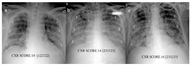
Imaging plays an important role in the detection of coronavirus (COVID-19) pneumonia in both managing the disease and evaluating the complications. Imaging with chest computed tomography (CT) can also have a potential predictive and prognostic role in COVID-19 patient outcomes. The aim of this pictorial review is to describe the role of imaging with chest X-ray (CXR), lung ultrasound (LUS), and CT in the diagnosis and management of COVID-19 pneumonia, the current indications, the scores proposed for each modality, the advantages/limitations of each modality and their role in detecting complications, and the histopathological correlations.

Keywords: ARDS; COVID-19; COVID-19 complications; COVID-19 pneumonia imaging guidelines; chest CT; chest CT protocols; chest CT severity scores; chest X-ray; chest X-ray protocols; chest X-ray scoring system; histopathological correlations; lung ultrasound; lung ultrasound protocols; lung ultrasound scoring system.


Conflict of interest statement

The authors do not report any conflicts of interest.

Figures

Figure 1
Figure 1
(a) Chest X-ray (CXR) of COVID-19 patient at bedside on anterior-posterior projection shows diffuse linear opacities associated with ground-glass opacity (GGO). (b) CXR of patient admitted to intensive care unit (ICU) showing two central venous catheters in superior vena cava (black arrows) and endotracheal tube (white arrow). Diffuse confluent GGO with consolidation in left lung and linear opacity and GGO in right lung are also visible.
Figure 2
Figure 2
CXR scores based on scores proposed by Maroldi et al. [27] in COVID-19 pneumonia. (a) Baseline score of 10 on CXR at bedside in AP projection at admission; (b) increased score of 14 reveals progression of COVID pneumonia on CXR follow-up on day 13; (c) score of 16 indicates further progression on day 18.
Figure 3
Figure 3
(a) Horizontal A line artifacts (solid white arrow) as regular aeration in a healthy lung, and normal pleural line (dashed white arrow); (b) B line vertical artifact (black arrow) in healthy lung with linear probe, and normal pleural line (dashed white arrow); (c) multiple B line artifacts (black arrow) with diffuse pleural thickness (dashed white arrow) in a patient with fibrosis. (Images courtesy of Dr. Luigi Monaco, head of the ultrasound unit at San Giuseppe Moscati Hospital.)
Figure 4
Figure 4
In this image is represented an example of the anatomical subdivision of the chest by 3 lines (median: parasternal line PSL; lateral: anterior axillary lines AAL; posterior: posterior axillary lines PAL) into 8 zones for the LUS examination.
Figure 5
Figure 5
Examples of lung ultrasound score (LUSS) based on score proposed by Dargent et al. [61] for patients with COVID-19 pneumonia. (a) LUS describes horizontal A lines for regular aeration (score: 0); (b) multiple B lines arising from thickened pleural line for moderate loss of aeration (1—B1); (c) coalescent B lines or white lung for severe loss of aeration (2—B2); (d) consolidation, with air bronchogram, for absence of aeration (3) and pleural effusion (white arrow).
Figure 6
Figure 6
(a) Lung embolisms in left pulmonary artery (white arrow) and right pulmonary artery (gray arrow); (b) extensive lung pulmonary embolism in left pulmonary artery, in lobar branch of inferior lobes (white arrow), and in lobar and segmentary branches of right inferior lobe (gray arrow).
Figure 7
Figure 7
(a) GGO pattern in COVID-19 pneumonia with peripheral and central distribution; (b) crazy paving pattern on right side with peripheral and posterior distribution; (c) consolidation areas in inferior lobes; (d) diffuse vascular enlargement (black arrow); (e) pleural thickness (white arrow); and (f) bronchiectasis (black arrow).
Figure 8
Figure 8
(a) Reverse halo sign (black arrow) consisting of central ground-glass opacity surrounded by complete ring of consolidation; (b) GGO areas surrounded by small consolidative area as halo sign (white arrow).
Figure 9
Figure 9
(a) Initial GGO pattern of COVID-19 pneumonia involving right superior lobe with peripheral distribution; (b) some GGO areas with interstitial thickness in both inferior lobes; (c,d) same areas in (a,b) evolving in consolidations.
Figure 10
Figure 10
Example of lung involvement in COVID-19 pneumonia based on CT-SS proposed by Pan et al. [108]. (a) GGO pattern in right (R) superior lobe with parenchymal involvement of <5%; (b) GGO in right superior lobe with parenchymal involvement of 5–25%; (c) GGO in right superior lobe with parenchymal involvement of 26–49%; (d) GGO with parenchymal involvement of 50–75% in right superior lobe and >75% in left (L) superior lobe.
Figure 11
Figure 11
(a) COVID-19 pneumonia on initial examination of an obese young patient with severe lung involvement (CT-SS of 18) that (b) further progressed to acute respiratory distress syndrome (ARDS) with diffuse consolidations.
Figure 12
Figure 12
Example of quantitative method using Thoracic-VCAR software that evaluates percentages of GGO, consolidations, and pulmonary parenchyma without COVID-19 pneumonia involvement.
Figure 13
Figure 13
(a) Diffuse subcutaneous emphysema and pneumomediastinum in a COVID-19 patient who underwent mechanical ventilation; (b) pneumopericardium (black arrow).

References

    1. Apolone G., Montomoli E., Manenti A., Boeri M., Sabia F., Hyseni I., Mazzini L., Martinuzzi D., Cantone L., Milanese G., et al. Unexpected detection of SARS-CoV-2 antibodies in the prepandemic period in Italy. Tumori J. 2020 doi: 10.1177/0300891620974755. - DOI - PMC - PubMed
    1. Deslandes A., Berti V., Tandjaoui-Lambotte Y., Alloui C., Carbonnelle E., Zahar J.R., Brichlera S., Cohen Y. SARS-COV-2 was already spreading in France in late December 2019. Int. J. Antimicrob. Agents. 2020;55:106006. doi: 10.1016/j.ijantimicag.2020.106006. - DOI - PMC - PubMed
    1. Stawicki S.P., Jeanmonod R., Miller A.C., Paladino L., Gaieski D.F., Yaffee A.Q., De Wulf A., Grover J., Papadimos T.J., Bloem C., et al. The 2019–2020 novel coronavirus (severe acute respiratory syndrome coronavirus 2) pandemic: A joint american college of academic international medicine-world academic council of emergency medicine multidisciplinary COVID-19 working group consensus paper. J. Glob. Infect. Dis. 2020;12:47–93. doi: 10.4103/jgid.jgid_86_20. - DOI - PMC - PubMed
    1. Mao R., Qiu Y., He J.-S., Tan J.-Y., Li X.-H., Liang J., Shen J., Zhu L.-R., Chen Y., Iacucci M., et al. Manifestations and prognosis of gastrointestinal and liver involvement in patients with COVID-19: A systematic review and meta-analysis. Lancet Gastroenterol. Hepatol. 2020;5:667–678. doi: 10.1016/S2468-1253(20)30126-6. - DOI - PMC - PubMed
    1. Gu J., Han B., Wang J. COVID-19: Gastrointestinal Manifestations and Potential Fecal–Oral Transmission. Gastroenterology. 2020;158:1518–1519. doi: 10.1053/j.gastro.2020.02.054. - DOI - PMC - PubMed

LinkOut - more resources