The Role of Transthoracic Ultrasound in the Study of Interstitial Lung Diseases: High-Resolution Computed Tomography Versus Ultrasound Patterns: Our Preliminary Experience
- PMID: 33806439
- PMCID: PMC8001146
- DOI: 10.3390/diagnostics11030439
The Role of Transthoracic Ultrasound in the Study of Interstitial Lung Diseases: High-Resolution Computed Tomography Versus Ultrasound Patterns: Our Preliminary Experience
Abstract
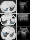
Transthoracic ultrasound (TUS) is a readily available imaging tool that can provide a quick real-time evaluation. The aim of this preliminary study was to establish a complementary role for this imaging method in the approach of interstitial lung diseases (ILDs). TUS examination was performed in 43 consecutive patients with pulmonary fibrosis and TUS findings were compared with the corresponding high-resolution computed tomography (HRCT) scans. All patients showed a thickened hyperechoic pleural line, despite no difference between dominant HRCT patterns (ground glass, honeycombing, mixed pattern) being recorded (p > 0.05). However, pleural lines' thickening showed a significant difference between different HRCT degree of fibrosis (p < 0.001) and a negative correlation with functional parameters. The presence of >3 B-lines and subpleural nodules was also assessed in a large number of patients, although they did not demonstrate any particular association with a specific HRCT finding or fibrotic degree. Results allow us to suggest a complementary role for TUS in facilitating an early diagnosis of ILD or helping to detect a possible disease progression or eventual complications during routine clinical practice (with pleural line measurements and subpleural nodules), although HRCT remains the gold standard in the definition of ILD pattern, disease extent and follow-up.
Keywords: high-resolution computed tomography; hyperechoic pleural line; interstitial lung diseases; screening tool; transthoracic ultrasound.
Conflict of interest statement
The authors declare no conflict of interest.
Figures


References
-
- Lee S.H., Park M.S., Kim S.Y., Kim D.S., Kim Y.W., Chung M.P., Uh S.T., Park C.S., Park S.W., Jeong S.H., et al. Factors affecting treatment outcome in patients with idiopathic nonspecific interstitial pneumonia: A nationwide cohort study. Respir. Res. 2017;18:204–212. doi: 10.1186/s12931-017-0686-7. - DOI - PMC - PubMed
-
- Travis W.D., Costabel U., Hansell D.M., King T.E., Jr., Lynch D.A., Nicholson A.G., Ryerson C.J., Ryu J.H., Selman M., Wells A.U., et al. An official American Thoracic Society/European Respiratory Society statement: Update of the international multidisciplinary classification of the idiopathic interstitial pneumonias. Am. J. Respir. Crit. Care Med. 2013;188:733–748. doi: 10.1164/rccm.201308-1483ST. - DOI - PMC - PubMed
-
- Raghu G., Remy-Jardin M., Myers J.L., Richeldi L., Ryerson C.J., Lederer D.J., Behr J., Cottin V., Danoff S.K., Morell F., et al. American Thoracic Society, European Respiratory Society, Japanese Respiratory Society, and Latin American Thoracic Society. Diagnosis of Idiopathic Pulmonary Fibrosis. An Official ATS/ERS/JRS/ALAT Clinical Practice Guideline. Am. J. Respir. Crit. Care Med. 2018;198:e44–e68. doi: 10.1164/rccm.201807-1255ST. - DOI - PubMed
-
- Buda N., Kosiak W., Wełnicki M., Skoczylas A., Olszewski R., Piotrkowski J., Skoczyński S., Radzikowska E., Jassem E., Grabczak E.M., et al. Recommendations for Lung Ultrasound in Internal Medicine. Recommendations for Lung Ultrasound in Internal Medicine. Diagnostics. 2020;10:597–621. doi: 10.3390/diagnostics10080597. - DOI - PMC - PubMed
LinkOut - more resources
Full Text Sources
Other Literature Sources

