. 2021 Mar 4;11(3):323.
doi: 10.3390/brainsci11030323.

Changes in Representation of Thalamic Projection Neurons within Prefrontal-Thalamic-Hippocampal Circuitry in a Rat Model of Third Trimester Binge Drinking

Affiliations

Changes in Representation of Thalamic Projection Neurons within Prefrontal-Thalamic-Hippocampal Circuitry in a Rat Model of Third Trimester Binge Drinking

Zachary H Gursky et al. Brain Sci. .

Abstract

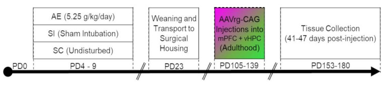
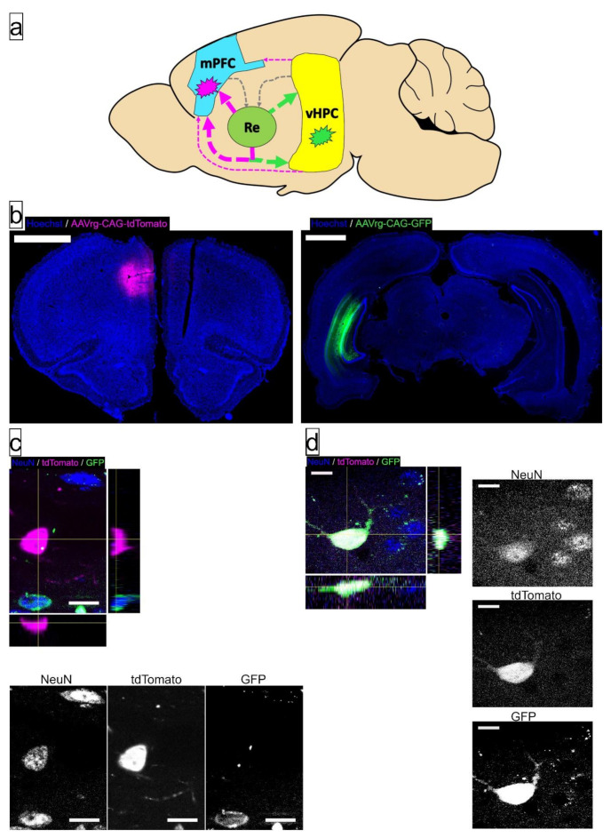
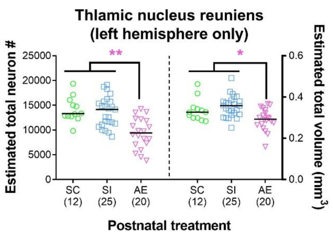
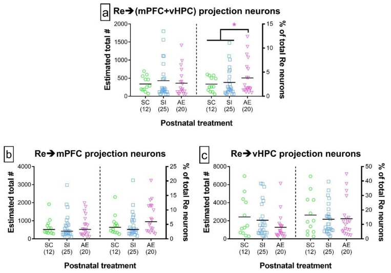
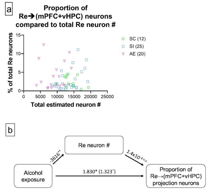
Alcohol exposure (AE) during the third trimester of pregnancy-a period known as the brain growth spurt (BGS)-could result in a diagnosis of a fetal alcohol spectrum disorder (FASD), a hallmark of which is impaired executive functioning (EF). Coordinated activity between prefrontal cortex and hippocampus is necessary for EF and thalamic nucleus reuniens (Re), which is required for prefrontal-hippocampal coordination, is damaged following high-dose AE during the BGS. The current experiment utilized high-dose AE (5.25 g/kg/day) during the BGS (i.e., postnatal days 4-9) of Long-Evans rat pups. AE reduces the number of neurons in Re into adulthood and selectively alters the proportion of Re neurons that simultaneously innervate both medial prefrontal cortex (mPFC) and ventral hippocampus (vHPC). The AE-induced change unique to Re→(mPFC + vHPC) projection neurons (neuron populations that innervate either mPFC or vHPC individually were unchanged) is not mediated by reduction in neuron number. These data are the first to examine mPFC-Re-HPC circuit connectivity in a rodent model of FASD, and suggest that both short-term AE-induced neuron loss and long-term changes in thalamic connectivity may be two distinct (but synergistic) mechanisms by which developmental AE can alter mPFC-Re-vHPC circuitry and impair EF throughout the lifespan.

Keywords: AAV; alcohol; connectivity; development; fetal alcohol spectrum disorders (FASD); immunofluorescence; immunohistochemistry; thalamus; unbiased stereology.


Conflict of interest statement

The authors declare no conflict of interest. The funders had no role in the design of the study; in the collection, analyses, or interpretation of data; in the writing of the manuscript, or in the decision to publish the results.

Figures

Figure 1
Figure 1
Experimental timeline for the current study.
Figure 2
Figure 2
Schematic representation and visualization of retrograde-labeling AAVrg-CAG injections into medial prefrontal cortex (mPFC), ventral hippocampus (HPC), and exemplar labeled cells in thalamic nucleus reuniens (Re). (a) A schematic indicating the approximate sites of AAV injection (indicated by magenta and green explosion symbol). Connectivity within mPFC-Re-HPC circuitry is presented as arrows, with the large, colored, arrows leaving Re as the population of projection neurons examined. Pairing of injection site and fluorophore was counterbalanced between postnatal treatment and sex. Connectivity diagram is derived from [48,49]; (b) Representative sections containing the center of the injection site into mPFC (left) and HPC (right). Scale bars = 2 mm; (c) Examples of Re projection neurons (NeuN+) projecting to the injection site of AAVrg-CAG-tdTomato (NeuN+/GFP/tdTomato+, in crosshairs), AAV-CAG-GFP (NeuN+/GFP+/tdTomato+, bottom left), or neither injection site (NeuN+/GFP/tdTomato, top right). Bars on right and bottom of image show Y-Z and X-Z planes of image stack, respectively, while the panels below show each color channel in isolation. Scale bar = 15 µm; (d) A subpopulation of neurons in Re project to both mPFC and HPC, as indicated by expression of fluorophores from both injection sites (NeuN+/GFP+/tdTomato+, in crosshairs). Bars on right and bottom of image show Y-Z and X-Z planes of image stack, respectively, while the panels on the right show each color channel in isolation. Scale bars = 15 µm.
Figure 3
Figure 3
High-dose alcohol exposure (AE) during the brain growth spurt (BGS) results in reduced number (#) of neurons in thalamic nucleus reuniens (Re) and a reduction in the total volume of Re. The AE postnatal treatment group had significantly lower number of neurons in Re (left) and significantly reduced total volume of Re (right), consistent with past studies [21,22]. Individual data points represent one animal and are superimposed over bars representing mean within that postnatal treatment. Data include both male and female animals, which are not differentiated due to the lack of sex effects. Sample sizes are given in parentheses below each treatment group. * p ≤ 0.050, ** p ≤ 0.010. For exact statistical values, see Table 3.
Figure 4
Figure 4
Quantification of Re projection neurons that simultaneously innervate mPFC and/or vHPC. (ac) Total number of Re projection neurons (left of each panel) and the proportion of total Re neurons comprised of this class of projection neuron (right of each panel) that innervated mPFC and vHPC concurrently (a), mPFC alone (b), or vHPC alone (c). While the total number of each class of projection neuron was not influenced by postnatal treatment, the proportion of Re→(mPFC + vHPC) neurons (relative to total neurons in Re) was significantly increased in the AE treatment group. For all panels in this figure, individual data points represent one animal and are superimposed over bars representing mean within that postnatal treatment. Data include both male and female animals, which are not differentiated due to the lack of sex effects. Sample sizes are given in parentheses below each treatment group. * p ≤ 0.050. For exact statistical values, see Table 4, Table 5 and Table 6 (and Tables S6–S8 for injection parameter-controlled values).
Figure 5
Figure 5
Neuron loss did not mediate the AE-induced increase in proportion of Re neurons that concurrently innervate mPFC and vHPC. (a) Scatterplot showing the relationship between total neuron number in Re and proportion of Re→(mPFC + vHPC) projection neurons; (b) Schematic representation of the mediation analysis between AE, Re neuron number, and proportion of Re→(mPFC + vHPC) projection neurons. The total effect of AE (in % of neurons) is shown in parentheses, while the direct effect is listed immediately next to the total effect (also in % of neurons). While the relationship between AE and Re neuron number was significant (estimate given in number of neurons), there was no significant association between Re neuron number and proportion of Re→(mPFC + vHPC) projection neurons, resulting in a lack of mediating effect. Data include both male and female animals, which are not differentiated due to the lack of sex effects. Sample sizes are given in parentheses next to each treatment group in the panel (a) legend. * p ≤ 0.050, ** p ≤ 0.010, n.s. = not statistically significant. For exact statistical values, see Section 3.3.

Similar articles

Cited by

References

    1. Burton R., Sheron N. No level of alcohol consumption improves health. Lancet. 2018;392:987–988. doi: 10.1016/S0140-6736(18)31571-X. - DOI - PubMed
    1. Bilbo S.D., Schwarz J.M. The immune system and developmental programming of brain and behavior. Front. Neuroendocr. 2012;33:267–286. doi: 10.1016/j.yfrne.2012.08.006. - DOI - PMC - PubMed
    1. Baram T.Z., Donato F., Holmes G.L. Construction and disruption of spatial memory networks during development. Learn. Mem. 2019;26:206–218. doi: 10.1101/lm.049239.118. - DOI - PMC - PubMed
    1. Klintsova A.Y., Hamilton G.F., Boschen K.E. Long-Term Consequences of Developmental Alcohol Exposure on Brain Structure and Function: Therapeutic Benefits of Physical Activity. Brain Sci. 2012;3:1–38. doi: 10.3390/brainsci3010001. - DOI - PMC - PubMed
    1. Astley S.J., Clarren S.K. Diagnosing the full spectrum of fetal alcohol-exposed individuals: introducing the 4-digit diagnostic code. Alcohol Alcohol. 2000;35:400–410. doi: 10.1093/alcalc/35.4.400. - DOI - PubMed

LinkOut - more resources