

Molecular Profiling and Gene Banking of Rabbit EPCs Derived from Two Biological Sources
- PMID: 33806502
- PMCID: PMC7998175
- DOI: 10.3390/genes12030366
Molecular Profiling and Gene Banking of Rabbit EPCs Derived from Two Biological Sources
Abstract
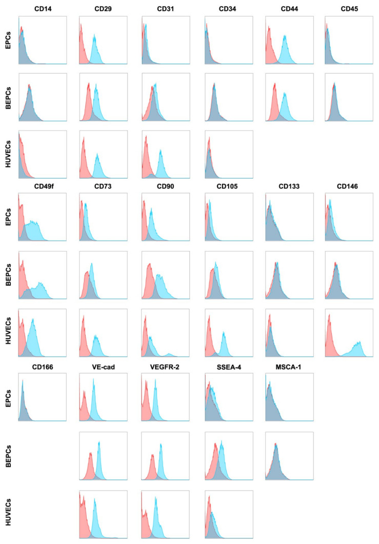
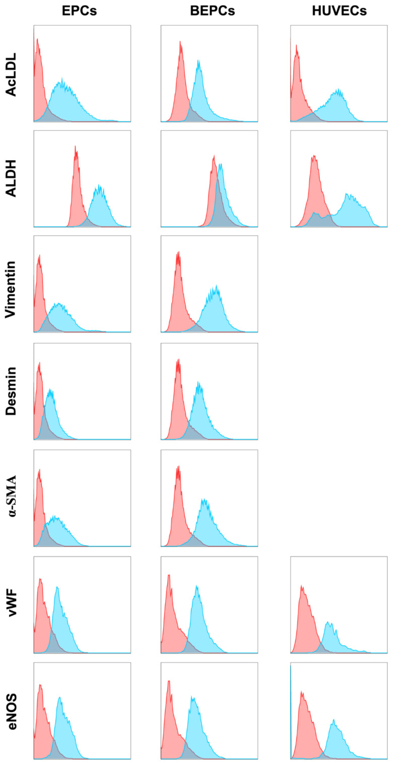
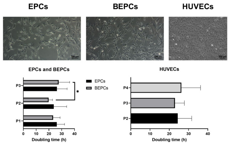
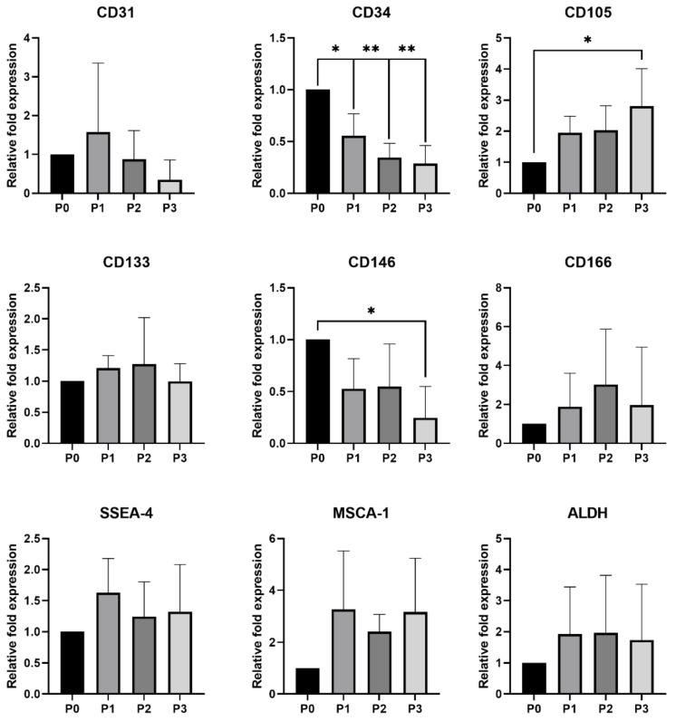
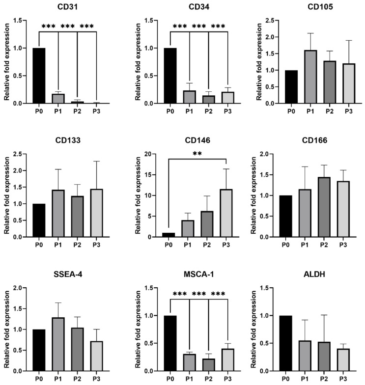
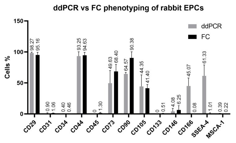
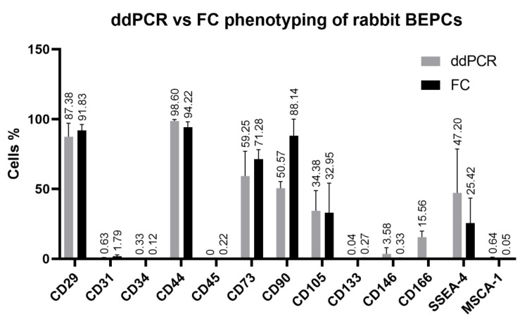
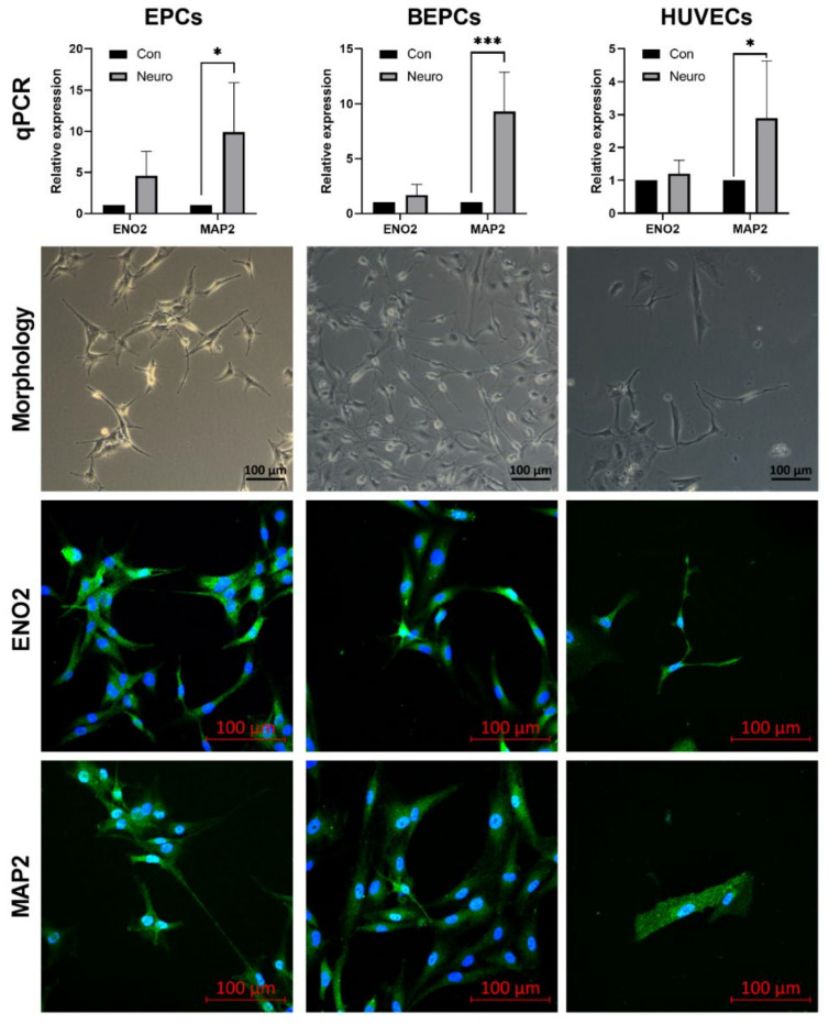
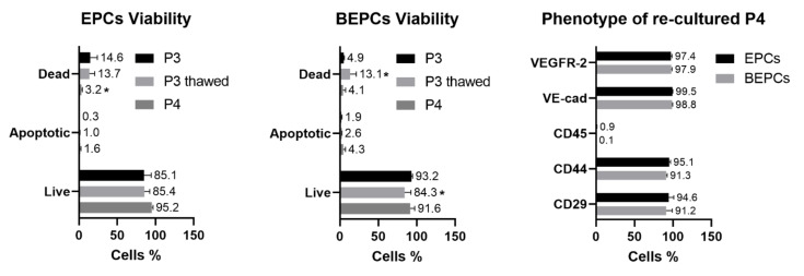
Endothelial progenitor cells (EPCs) have been broadly studied for several years due to their outstanding regenerative potential. Moreover, these cells might be a valuable source of genetic information for the preservation of endangered animal species. However, a controversy regarding their characterization still exists. The aim of this study was to isolate and compare the rabbit peripheral blood- and bone marrow-derived EPCs with human umbilical vein endothelial cells (HUVECs) in terms of their phenotype and morphology that could be affected by the passage number or cryopreservation as well as to assess their possible neuro-differentiation potential. Briefly, cells were isolated and cultured under standard endothelial conditions until passage 3. The morphological changes during the culture were monitored and each passage was analyzed for the typical phenotype using flow cytometry, quantitative real-time polymerase chain reaction (qPCR) and novel digital droplet PCR (ddPCR), and compared to HUVECs. The neurogenic differentiation was induced using a commercial kit. Rabbit cells were also cryopreserved for at least 3 months and then analyzed after thawing. According to the obtained results, both rabbit EPCs exhibit a spindle-shaped morphology and high proliferation rate. The both cell lines possess same stable phenotype: CD14-CD29+CD31-CD34-CD44+CD45-CD49f+CD73+CD90+CD105+CD133-CD146-CD166+VE-cadherin+VEGFR-2+SSEA-4+MSCA-1-vWF+eNOS+AcLDL+ALDH+vimentin+desmin+α-SMA+, slightly different from HUVECs. Moreover, both induced rabbit EPCs exhibit neuron-like morphological changes and expression of neuronal markers ENO2 and MAP2. In addition, cryopreserved rabbit cells maintained high viability (>85%) and endothelial phenotype after thawing. In conclusion, our findings suggest that cells expanded from the rabbit peripheral blood and bone marrow are of the endothelial origin with a stable marker expression and interesting proliferation and differentiation capacity.
Keywords: HUVECs; bone marrow; cryopreservation; ddPCR; endothelial progenitor cells; flow cytometry; neuro-transdifferentiation; peripheral blood; qPCR; rabbit.
Conflict of interest statement
The authors declare no conflict of interest.
Figures
Similar articles
-
Combination of granulocyte colony-stimulating factor and CXCR4 antagonist AMD3100 for effective harvest of endothelial progenitor cells from peripheral blood and in vitro formation of primitive endothelial networks.Cell Tissue Bank. 2016 Mar;17(1):161-9. doi: 10.1007/s10561-015-9527-4. Epub 2015 Jul 31. Cell Tissue Bank. 2016. PMID: 26224208
-
Combined approach for characterization and quality assessment of rabbit bone marrow-derived mesenchymal stem cells intended for gene banking.N Biotechnol. 2020 Jan 25;54:1-12. doi: 10.1016/j.nbt.2019.08.001. Epub 2019 Aug 7. N Biotechnol. 2020. PMID: 31400479
-
Determine exogenous human DDAH2 gene function in rabbit bone marrow-derived endothelial progenitor cells in vitro.Cell Biochem Funct. 2017 Mar;35(2):69-76. doi: 10.1002/cbf.3249. Epub 2017 Feb 1. Cell Biochem Funct. 2017. PMID: 28150318
-
Phenotypical Characterization and Neurogenic Differentiation of Rabbit Adipose Tissue-Derived Mesenchymal Stem Cells.Genes (Basel). 2021 Mar 17;12(3):431. doi: 10.3390/genes12030431. Genes (Basel). 2021. PMID: 33802902 Free PMC article.
-
Update on the pathogenesis of Scleroderma: focus on circulating progenitor cells.F1000Res. 2016 Apr 22;5:F1000 Faculty Rev-723. doi: 10.12688/f1000research.7986.1. eCollection 2016. F1000Res. 2016. PMID: 27158466 Free PMC article. Review.
Cited by
-
Secretome Analysis of Rabbit and Human Mesenchymal Stem and Endothelial Progenitor Cells: A Comparative Study.Int J Mol Sci. 2021 Nov 13;22(22):12283. doi: 10.3390/ijms222212283. Int J Mol Sci. 2021. PMID: 34830165 Free PMC article.
-
Changes in the Intracellular Composition of Macro and Microminerals After Cryopreservation of the Rabbit Stem/Progenitor Cells.J Dev Biol. 2025 Feb 21;13(1):6. doi: 10.3390/jdb13010006. J Dev Biol. 2025. PMID: 40137013 Free PMC article.
-
Role of Bushen Huoxue Formula and transplanted endothelial progenitor cells play in promoting endplate microcirculation and attenuating intervertebral disc degeneration.Heliyon. 2024 Mar 19;10(6):e28095. doi: 10.1016/j.heliyon.2024.e28095. eCollection 2024 Mar 30. Heliyon. 2024. PMID: 38545187 Free PMC article.
-
Therapeutic Potential of Mesenchymal Stem Cell and Tenocyte Secretomes for Tendon Repair: Proteomic Profiling and Functional Characterization In Vitro and In Ovo.Int J Mol Sci. 2025 Apr 11;26(8):3622. doi: 10.3390/ijms26083622. Int J Mol Sci. 2025. PMID: 40332130 Free PMC article.
-
Generation of Novel Monoclonal Antibodies Recognizing Rabbit CD34 Antigen.Biomolecules. 2025 Jul 15;15(7):1021. doi: 10.3390/biom15071021. Biomolecules. 2025. PMID: 40723893 Free PMC article.
References
-
- Vašíček J., Kováč M., Baláži A., Kulíková B., Tomková M., Olexíková L., Čurlej J., Bauer M., Schnabl S., Hilgarth M., et al. Combined approach for characterization and quality assessment of rabbit bone marrow-derived mesenchymal stem cells intended for gene banking. New Biotechnol. 2020;54:1–12. doi: 10.1016/j.nbt.2019.08.001. - DOI - PubMed
Publication types
MeSH terms
Substances
LinkOut - more resources
Full Text Sources
Other Literature Sources
Research Materials
Miscellaneous
