

Vitamin D Status, Bone Mineral Density, and VDR Gene Polymorphism in a Cohort of Belarusian Postmenopausal Women
- PMID: 33806559
- PMCID: PMC7999336
- DOI: 10.3390/nu13030837
Vitamin D Status, Bone Mineral Density, and VDR Gene Polymorphism in a Cohort of Belarusian Postmenopausal Women
Abstract
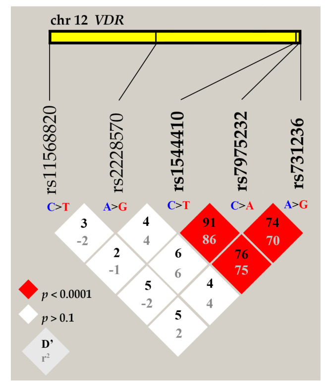
Vitamin D plays an important role in bone metabolism and is important for the prevention of multifactorial pathologies, including osteoporosis (OP). The biological action of vitamin is realized through its receptor, which is coded by the VDR gene. VDR gene polymorphism can influence individual predisposition to OP and response to vitamin D supplementation. The aim of this work was to reveal the effects of VDR gene ApaI rs7975232, BsmI rs1544410, TaqI rs731236, FokI rs2228570, and Cdx2 rs11568820 variants on bone mineral density (BMD), 25-hydroxyvitamin D level, and OP risk in Belarusian women.
Methods: The case group included 355 women with postmenopausal OP, and the control group comprised 247 women who met the inclusion criteria. TaqMan genotyping assay was used to determine VDR gene variants.
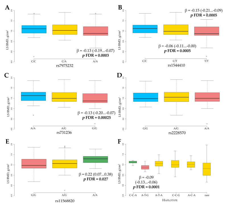
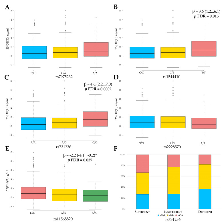
Results: Rs7975232 A/A, rs1544410 T/T, and rs731236 G/G single variants and their A-T-G haplotype showed a significant association with increased OP risk (for A-T-G, OR = 1.8, p = 0.0001) and decreased BMD (A-T-G, -0.09 g/cm2, p = 0.0001). The rs11568820 A-allele showed a protective effect on BMD (+0.22 g/cm2, p = 0.027). A significant dose effect with 25(OH)D was found for rs1544410, rs731236, and rs11568820 genotypes. Rs731236 A/A was associated with the 25(OH)D deficiency state.
Conclusion: Our novel data on the relationship between VDR gene variants and BMD, 25(OH)D level, and OP risk highlights the importance of genetic markers for personalized medicine strategy.
Keywords: VDR gene; bone mineral density; osteoporosis; polymorphism; predisposition; vitamin D.
Conflict of interest statement
The authors declare no conflict of interest. The funders had no role in the design of the study; in the collection, analyses, or interpretation of data; in the writing of the manuscript, or in the decision to publish the results.
Figures
Similar articles
-
Relationship Between Vitamin D Receptor Gene BsmI Polymorphism and 25-Hydroxyvitamin D Total Levels in Slovak Postmenopausal Women with Reduced Bone Mineral Density.Genes (Basel). 2025 Mar 13;16(3):337. doi: 10.3390/genes16030337. Genes (Basel). 2025. PMID: 40149488 Free PMC article.
-
Serum 25(OH)D concentration, common variants of the VDR gene and lung cancer occurrence.Int J Cancer. 2017 Jul 15;141(2):336-341. doi: 10.1002/ijc.30740. Epub 2017 Apr 24. Int J Cancer. 2017. PMID: 28411367
-
Association analysis of vitamin D receptor gene polymorphisms and bone mineral density in postmenopausal Mexican-Mestizo women.Genet Mol Res. 2013 Jul 30;12(3):2755-63. doi: 10.4238/2013.July.30.13. Genet Mol Res. 2013. PMID: 23979900
-
Vitamin D status and the Cdx-2 polymorphism of the vitamin D receptor gene are determining factors of bone mineral density in young healthy postmenopausal women.J Steroid Biochem Mol Biol. 2013 Jul;136:187-9. doi: 10.1016/j.jsbmb.2012.09.026. Epub 2012 Sep 28. J Steroid Biochem Mol Biol. 2013. PMID: 23026509 Review.
-
Associations between VDR Gene Polymorphisms and Osteoporosis Risk and Bone Mineral Density in Postmenopausal Women: A systematic review and Meta-Analysis.Sci Rep. 2018 Jan 17;8(1):981. doi: 10.1038/s41598-017-18670-7. Sci Rep. 2018. Retraction in: Sci Rep. 2021 Apr 21;11(1):9030. doi: 10.1038/s41598-021-88654-1. PMID: 29343720 Free PMC article. Retracted.
Cited by
-
Research progress on mesenchymal stem cell‑derived exosomes in the treatment of osteoporosis induced by knee osteoarthritis (Review).Int J Mol Med. 2025 Oct;56(4):160. doi: 10.3892/ijmm.2025.5601. Epub 2025 Aug 1. Int J Mol Med. 2025. PMID: 40747674 Free PMC article. Review.
-
The VDR rs1544410 and rs11568820 Variants and the Risk of Osteoporosis in the Polish Population.Int J Mol Sci. 2025 Jan 8;26(2):481. doi: 10.3390/ijms26020481. Int J Mol Sci. 2025. PMID: 39859195 Free PMC article.
-
Impact of Vitamin D on Osseointegration in Dental Implants: A Systematic Review of Human Studies.Nutrients. 2024 Jan 9;16(2):209. doi: 10.3390/nu16020209. Nutrients. 2024. PMID: 38257102 Free PMC article.
-
Identification of Immune Hub Genes in Obese Postmenopausal Women Using Microarray and Single-Cell RNA Seq Data.Genes (Basel). 2025 Jun 30;16(7):783. doi: 10.3390/genes16070783. Genes (Basel). 2025. PMID: 40725440 Free PMC article.
-
Relationship Between Vitamin D Receptor Gene BsmI Polymorphism and 25-Hydroxyvitamin D Total Levels in Slovak Postmenopausal Women with Reduced Bone Mineral Density.Genes (Basel). 2025 Mar 13;16(3):337. doi: 10.3390/genes16030337. Genes (Basel). 2025. PMID: 40149488 Free PMC article.
References
-
- Hernlund E., Svedbom A., Ivergård M., Compston J., Cooper C., Stenmark J., McCloskey E.V., Jönsson B., Kanis J.A. Osteoporosis in the European Union: Medical management, epidemiology and economic burden. A report prepared in collaboration with the International Osteoporosis Foundation (IOF) and the European Federation of Pharmaceutical Industry Associations (EFPIA) Arch. Osteoporos. 2013;8:136. doi: 10.1007/s11657-013-0136-1. - DOI - PMC - PubMed
MeSH terms
Substances
Grants and funding
LinkOut - more resources
Full Text Sources
Other Literature Sources
Medical
Miscellaneous
