Review
. 2021 Mar 4;11(3):382.
doi: 10.3390/biom11030382.

Interfaces with Structure Dynamics of the Workhorses from Cells Revealed through Cross-Linking Mass Spectrometry (CLMS)

Affiliations
Review

Interfaces with Structure Dynamics of the Workhorses from Cells Revealed through Cross-Linking Mass Spectrometry (CLMS)

Umesh Kalathiya et al. Biomolecules. .

Abstract

The fundamentals of how protein-protein/RNA/DNA interactions influence the structures and functions of the workhorses from the cells have been well documented in the 20th century. A diverse set of methods exist to determine such interactions between different components, particularly, the mass spectrometry (MS) methods, with its advanced instrumentation, has become a significant approach to analyze a diverse range of biomolecules, as well as bring insights to their biomolecular processes. This review highlights the principal role of chemistry in MS-based structural proteomics approaches, with a particular focus on the chemical cross-linking of protein-protein/DNA/RNA complexes. In addition, we discuss different methods to prepare the cross-linked samples for MS analysis and tools to identify cross-linked peptides. Cross-linking mass spectrometry (CLMS) holds promise to identify interaction sites in larger and more complex biological systems. The typical CLMS workflow allows for the measurement of the proximity in three-dimensional space of amino acids, identifying proteins in direct contact with DNA or RNA, and it provides information on the folds of proteins as well as their topology in the complexes. Principal CLMS applications, its notable successes, as well as common pipelines that bridge proteomics, molecular biology, structural systems biology, and interactomics are outlined.

Keywords: CLMS; chemical cross-linkers; cross-linking mass spectrometry; protein–DNA; protein–RNA interactions; protein–protein; proteomics; structural biology.


Conflict of interest statement

The authors declare no conflict of interest.

Figures

Figure 1
Figure 1
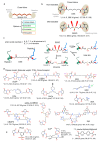
A spectrum of widely characterized experimental methods, based on biochemical, biophysical, or genetic principles. The listed methods define protein–protein interactions at various degrees of affinity as well as specificity [1,2,11,12,13,19,20,21,22,23,24,25,26,27,28,29,30,31,32,33,34,35,36,37,38,39,40]. The RNA and DNA diagrams, representing the genetic methods, were prepared using the BIOVIA draw (Dassault Systèmes, BIOVIA Corp., San Diego, CA, USA) tool.
Figure 4
Figure 4
Cross-linking reagents anatomy, chemistry, and evolution of cross-linkers. (a) Cross-linking reagents comprise various reactive groups, spacer scaffolds with different lengths. Based on the applied experimental workflow, a variety of isotope labeled, cleavable, enrichable, releasable, and spacer groups are in use. A large number of cross-linkers establish their combinatorial complexity and further supports a larger number of future cross-linkers [87,88,89]. (b,c) Structures of the commonly used cross-linkers for analyzing protein–protein interactions involving non-cleavable (disuccinimidyl suberate, DSS) and MS-cleavable (disuccinimidyl sulfoxide, DSSO) cross-linkers [71,91]. Representation of the suggested fragmentation schemes (interlink as well as intralink cross-linked peptides) of DSS and DSSO cross-linked peptides. (d) In addition to DSS and DSSO, most commonly used cross-linkers are shown (SuDP, Disuccinimidylsuccinamyl aspartyl proline; DSBU, Disuccinimidyldibutyric urea; BS3, Bis(sulfosuccinimidyl) suberate; DC4, 1,4-Bis{4-[(2,5-dioxo-1-pyrrolidinyl) oxy]-4-oxobutyl}-1,4-diazoniabicyclo[2.2.2]octaneoctane; PAC4, 1,1-Bis{4-[(2,5-dioxopyrrolidin-1-yl)oxy]-4-oxobutyl}-4-ethynylpiperidin-1-ium; EDC, 1-Ethyl-3-(3-dimethylaminopropyl)carbodiimide; Azide-A-DSBSO, Azide-tagged, acid-cleavable disuccinimidyl bissulfoxide; SDA, succinimidyl 4,4’-azipentanoate; CBDPS, Cyanurbiotindipropionyl succinimide; and PIR, protein interaction reporter) [71,72,89,91,92,93,94,95,96,97,98,99,100]. (e) For the enrichment of cross-linked products/peptides a few specific cross-linkers areused such as:PhoX linker (non-cleavable but containing a phosphonic group) [101], CLIP cross-linker (introduces alkyne groups to cross-linked peptides) [102], and 13C labeled form of biphenyldiglyoxal (diglyoxal cross-linkers) [103,104]. For the cross-linkers on panel b, d, and e, different properties such as spacer length, molecular weight, topological polar surface are (TPSA), and number of hydrogen bond donor or acceptor atoms in the cross linker are described. TPSA and hydrogen bonds were computed using the Molecular Operating Environment (MOE; Chemical Computing Group Inc., Montreal, QC, Canada) package, and the protein/peptide/chemical structures were prepared using BIOVIA Discovery Studio visualizer and BIOVIA draw tools (DassaultSystèmes, BIOVIA Corp., San Diego, CA, USA).
Figure 6
Figure 6
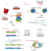
Schematic outline of the CLMS technique, to probe protein- DNA /RNA interactions. (a) Workflow for mass spectrometry analysis of UV-induced cross-links in histone-DNA complexes, nucleosomes, and chromatin arrays [140,159]. (b) Cross-linking based methods use either UV (RAP, RNA affinity purification; PAIR, peptide-nucleic acid assisted identification of RNA-binding proteins; MS2-BioTRAP, MS2 in vivo biotin-tagged RAP; and TRIP, tandem RNA isolation procedure) or formaldehyde cross-linking (CHART, capture hybridization analysis of RNA targets; ChIRP, Chromatin Isolation by RNA Purification) to capture RNA–protein interactions. Biotinylated oligonucleotide probes are hybridized to the RNA, and then the RNA and cross-linked proteins are purified for downstream analysis (RBP, RNA-binding protein; clRNPs, cross-linked RNPs) [149,160]. Representations of the protein or peptide structures in the figure, were prepared using BIOVIA Discovery Studio (Dassault Systèmes, BIOVIA Corp., San Diego, CA, USA) visualizer.
Figure 2
Figure 2
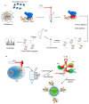
The generic workflow of cross-linking mass spectrometry (CLMS) experiments. (a) A wide range of samples (from purified proteins to intact tissues and organs) applicable to perform CLMS experiments [11,80]. (b) In a representative CLMS workflow, a selected linker is applied to the sample and the cross-linking reaction is carried out. Depending on the actual chemistry of interest, the reaction is stopped through chemical quenching or removal of the reagents. The proteins can be then digested in solution or gel to produce a mix of cross-linked and linear peptides. Prior to mass spectrometry analysis, the cross-linked peptides are often enriched by chromatographic methods, for example, the size exclusion chromatography, ion exchange chromatography, or purification through an affinity tag. Finally, the sample is subjected to LC-MS/MS (Liquid chromatography-tandem mass spectrometry) acquisition pipelines that have been developed to add the likelihood of selecting cross-linked peptide precursors for fragmentation. Using a variety of search software, two linked peptides can be identified from spectra and through the methods determining the false discovery rate, the list of matches can be filtered to the desired confidence. The cross-links can be also visualized by integrative modeling techniques. CLMS techniques convey the structural information in the form of distance restraints on single protein, protein complexes and allows to portray protein networks [11,80,81,82]. In this figure the structures of protein or peptides were prepared using BIOVIA Discovery Studio (Dassault Systèmes, BIOVIA Corp., San Diego, CA, USA) visualizer tool.
Figure 3
Figure 3
Three important mass spectrometry approaches used in the structural proteomics analysis of binary complexes. In the hydrogen/deuterium exchange, H2O is replaced by D2O and the resulting exchange associated with a mass increase can be detected by mass spectrometry (MS). Chemical cross-linking, comprises the covalent coupling of two reactive groups within a protein or between two different proteins, and then by introducing cross-links at the specific residues, spatial information at different levels can be obtained using MS. In covalent labeling, irreversible modifications are introduced at the reactive side chains and the solvent or surface exposed residues in proteins can be identified [23,72]. In this figure the protein or peptide structures were prepared using the BIOVIA Discovery Studio (Dassault Systèmes, BIOVIA Corp., San Diego, CA, USA) visualizer.
Figure 5
Figure 5
A workflow of determining protein structure, as well as the protein–protein interactions using the most frequent bottom-up cross-linking mass spectrometry. Extracted cross-linked proteins could be turned into peptides by several digestion protocols and proteases. Protein digestion protocol is chosen based upon up-stream steps taken to prepare cross-linked protein extract. FASP (filter aided sample preparation), ‘in-gel’, and ‘out-gel’ digestion protocols are used predominantly when a sample contains mass spectrometry incompatible substances, e.g., detergents or contaminants. However, ‘in-solution’ digestion might be used along with proteomic samples, which are already mass spectrometry compatible and contain predominantly cross-linked proteins of interest. Complexity of the cross-linked peptide samples might be reduced by separating cross-linked peptides from linear peptides after digestion. Difference in physico-chemical properties of cross-linked and linear peptides is often exploited to perform strong cation exchange or separation based on difference of peptide size. Alternatively, cross-linked peptides could be pulled down if a linker containing an affinity tag such as for example, biotin group has been used. Down-stream mass spectrometry analysis is frequently done in data-dependent acquisition mode (DDA). In the figure, representation of protein or peptide structures were prepared using BIOVIA Discovery Studio (Dassault Systèmes, BIOVIA Corp., San Diego, CA, USA) visualizer.

References

    1. Zhou M., Li Q., Wang R. Current experimental methods for characterizing protein-protein interactions. Chem. Med. Chem. 2016;11:738–756. doi: 10.1002/cmdc.201500495. - DOI - PMC - PubMed
    1. Berggård T., Linse S., James P. Methods for the detection and analysis of protein-protein interactions. Proteomics. 2007;16:2833–2842. doi: 10.1002/pmic.200700131. - DOI - PubMed
    1. Brown C.J., Lain S., Verma C.S., Fersht A.R., Lane D.P. Awakening guardian angels: Drugging the p53 pathway. Nat. Rev. Cancer. 2009;12:862–873. doi: 10.1038/nrc2763. - DOI - PubMed
    1. Joerger A.C., Fersht A.R. Structure-function-rescue: The diverse nature of common p53 cancer mutants. Oncogene. 2007;26:2226–2242. doi: 10.1038/sj.onc.1210291. - DOI - PubMed
    1. Brooke M.A., Nitoiu D., Kelsell D.P. Cell-cell connectivity: Desmosomes and disease. J. Pathol. 2012;226:158–171. doi: 10.1002/path.3027. - DOI - PubMed

Publication types

LinkOut - more resources