. 2021 Mar 4;13(3):187.
doi: 10.3390/toxins13030187.

Zearalenone Induces Endothelial Cell Apoptosis through Activation of a Cytosolic Ca2+/ERK1/2/p53/Caspase 3 Signaling Pathway

Affiliations

Zearalenone Induces Endothelial Cell Apoptosis through Activation of a Cytosolic Ca2+/ERK1/2/p53/Caspase 3 Signaling Pathway

Hyeon-Ju Lee et al. Toxins (Basel). .

Abstract

Zearalenone (ZEN) is a mycotoxin that has been reported to damage various types of cells/tissues, yet its effects on endothelial cells (ECs) have never been investigated. Therefore, this study investigates the potential effects of ZEN using bovine aortic ECs (BAECs). In this study, we found that ZEN induced apoptosis of BAECs through increased cleavage of caspase 3 and poly ADP-ribose polymerase (PARP). ZEN also increased phosphorylation of ERK1/2 and p53, and treatment with the ERK1/2 or p53 inhibitor reversed ZEN-induced EC apoptosis. Transfection of BAECs with small interfering RNA against ERK1/2 or p53 revealed ERK1/2 as an upstream target of p53 in ZEN-stimulated apoptosis. ZEN increased the production of reactive oxygen species (ROS), yet treatment with the antioxidant did not prevent EC apoptosis. Similarly, blocking of estrogen receptors by specific inhibitors also did not prevent ZEN-induced apoptosis. Finally, chelation of cytosolic calcium (Ca2+) using BAPTA-AM or inhibition of endoplasmic reticulum (ER) Ca2+ channel using 2-APB reversed ZEN-induced EC apoptosis, but not by inhibiting ER stress using 4-PBA. Together, our findings demonstrate that ZEN induces EC apoptosis through an ERK1/2/p53/caspase 3 signaling pathway activated by Ca2+ release from the ER, and this pathway is independent of ROS production and estrogen receptor activation.

Keywords: apoptosis; calcium; endothelial cells; mycotoxin; zearalenone.


Conflict of interest statement

All authors declare they have no conflict of interest.

Figures
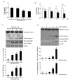
Figure 1
Figure 1
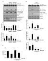
ZEN reduces the viability of BAECs by caspase-dependent apoptosis. BAECs were treated with various concentrations of ZEN (0, 10, 30 or 60 µM) for 24 h or 30 µM of ZEN for various time points (0, 4, 8, 16 or 24 h). (a,b) Cell viability was measured using the MTT assay. (c,d) The protein expression of cleaved caspase 3 and PARP in the BAECs was quantified (relative to relative to tubulin) using western blot analyses. (e) Apoptosis induced by ZEN at different concentrations was measured by FACS using annexin V/PI staining. After the pretreatment of 20 µM Z-DEVD-FMK for 1 h, BAECs were incubated with 30 µM ZEN for 24 h. (f) The protein expression of cleaved caspase 3 and PARP relative to tubulin was quantified using western blot analyses. (g) Apoptosis was measured by FACS using annexin V/PI staining. The plots depict the mean fold changes relative to the control (±SD) from at least four independent experimental trials. The different alphabetical letters refer to significant difference (p < 0.05) among groups, which were determined by one-way ANOVA followed by Tukey’s multiple comparisons.
Figure 1
Figure 1
ZEN reduces the viability of BAECs by caspase-dependent apoptosis. BAECs were treated with various concentrations of ZEN (0, 10, 30 or 60 µM) for 24 h or 30 µM of ZEN for various time points (0, 4, 8, 16 or 24 h). (a,b) Cell viability was measured using the MTT assay. (c,d) The protein expression of cleaved caspase 3 and PARP in the BAECs was quantified (relative to relative to tubulin) using western blot analyses. (e) Apoptosis induced by ZEN at different concentrations was measured by FACS using annexin V/PI staining. After the pretreatment of 20 µM Z-DEVD-FMK for 1 h, BAECs were incubated with 30 µM ZEN for 24 h. (f) The protein expression of cleaved caspase 3 and PARP relative to tubulin was quantified using western blot analyses. (g) Apoptosis was measured by FACS using annexin V/PI staining. The plots depict the mean fold changes relative to the control (±SD) from at least four independent experimental trials. The different alphabetical letters refer to significant difference (p < 0.05) among groups, which were determined by one-way ANOVA followed by Tukey’s multiple comparisons.
Figure 2
Figure 2
ERK1/2 mediates apoptosis by ZEN in BAECs. (a) The protein expression of cleaved caspase 3 and PARP relative to tubulin was quantified using western blot analyses in BAECs exposed to 30 µM of ZEN for 24 h after pretreatment with JNK inhibitor SP600125 (1 μM) or p38 MAPK inhibitor SB203580 (5 μM) for 1 h. SB203580 does not directly affect phosphorylation of p38, but inhibits p38 catalytic activity by binding to the ATP binding pocket, inhibiting phosphorylation of MAPKAPK, a downstream molecule of p38 MAPK. The plots are representative of at least four independent experimental trials. (b) After pretreatment with 1 µM of ERK1/2 inhibitor U0126 for 1 h, BAECs were incubated with 30 µM of ZEN for 24 h. The protein expression of cleaved caspase 3 and PARP, p-ERK1/2, and ERK1/2 relative to tubulin was quantified using western blot analyses. (c) Cell viability was measured using the MTT assay. The plots depict the mean fold changes relative to the control (±SD) from at least four independent experimental trials. The different alphabetical letters refer to significant difference (p < 0.05) among groups, which were determined by one-way ANOVA followed by Tukey’s multiple comparisons.
Figure 3
Figure 3
p53 is involved in EC apoptosis by ZEN. After pretreatment with 5 µM of p53 inhibitor pifithrin-α for 1 h, BAECs were incubated with 30 µM of ZEN for 24 h. (a) Cell viability was measured by using the MTT assay. (b) The protein expression of cleaved caspase 3 and PARP, and p-p53 relative to tubulin was quantified using western blot analyses. The plots depict the mean fold changes relative to the control (±SD) from at least four independent experimental trials. The different alphabetical letters refer to significant difference (p < 0.05) among groups, which were determined by one-way ANOVA followed by Tukey’s multiple comparisons.
Figure 4
Figure 4
ZEN-induced EC apoptosis is mediated via a signaling axis of ERK1/2/p53/caspase 3. (a) After pretreatment with 1 µM of U0126 or 5 µM of pifithrin-α for 1 h, BAECs were incubated with 30 µM of ZEN for 24 h, and the protein expression of p-p53 and p-ERK1/2 relative to tubulin was quantified using western blot analyses. The plots depict the mean fold changes relative to the control (±SD) from at least four independent experimental trials. (b,c) The protein expression of cleaved caspase 3 and PARP relative to tubulin were quantified using western blot analyses in the BAECs transfected with siRNA of ERK1/2 and p53 with or without 30 µM of ZEN exposure for 24 h. (d) The mRNA expression of p53 relative to GAPDH in the BAECs were quantified using RT-PCR analyses. The plots depict the mean fold changes relative to the control (±SD) from at least four independent experimental trials. The different alphabetical letters refer to significant difference (p < 0.05) among groups, which were determined by one-way ANOVA followed by Tukey’s multiple comparisons.
Figure 4
Figure 4
ZEN-induced EC apoptosis is mediated via a signaling axis of ERK1/2/p53/caspase 3. (a) After pretreatment with 1 µM of U0126 or 5 µM of pifithrin-α for 1 h, BAECs were incubated with 30 µM of ZEN for 24 h, and the protein expression of p-p53 and p-ERK1/2 relative to tubulin was quantified using western blot analyses. The plots depict the mean fold changes relative to the control (±SD) from at least four independent experimental trials. (b,c) The protein expression of cleaved caspase 3 and PARP relative to tubulin were quantified using western blot analyses in the BAECs transfected with siRNA of ERK1/2 and p53 with or without 30 µM of ZEN exposure for 24 h. (d) The mRNA expression of p53 relative to GAPDH in the BAECs were quantified using RT-PCR analyses. The plots depict the mean fold changes relative to the control (±SD) from at least four independent experimental trials. The different alphabetical letters refer to significant difference (p < 0.05) among groups, which were determined by one-way ANOVA followed by Tukey’s multiple comparisons.
Figure 5
Figure 5
Estrogen receptors do not mediate ECs apoptosis by ZEN. After pretreatment with (a,b) 10 µM of ICI 182,780 or (c,d) 1 µM of G-15 for 1 h, BAECs were incubated with 30 µM of ZEN for 24 h. (a,c) Cell viability was measured using the MTT assay. (b,d) The protein expression of cleaved caspase 3 and PARP relative to tubulin was quantified using western blot analyses. The plots depict the mean fold changes relative to the control (±SD) from at least four independent experimental trials. The different alphabetical letters refer to significant differences (p < 0.05) among groups, which were determined by one-way ANOVA followed by Tukey’s multiple comparisons.
Figure 6
Figure 6
ZEN induces EC apoptosis in an ROS-independent manner. After pretreatment with 5 mM of NAC for 3 h, BAECs were incubated with 30 µM of ZEN for 24 h. (a) The intracellular ROS production from the BAECs was measured using DCF-DA. (b) Cell viability was measured by using the MTT assay. (c) The protein expression of cleaved caspase 3 and PARP relative to tubulin was quantified using western blot analyses. The plots depict the mean fold changes relative to the control (±SD) from at least four independent experimental trials. The different alphabetical letters refer to significant difference (p < 0.05) among groups, which were determined by one-way ANOVA followed by Tukey’s multiple comparisons.
Figure 7
Figure 7
Cytosolic Ca2+ is involved in ZEN-induced EC apoptosis. After pretreatment with 2 µM of BAPTA-AM (ac) or 0.1 mM of EGTA (d) for 1 h, BAECs were incubated with 30 µM of ZEN for 24 h. (a) Cell viability was measured using the MTT assay. (b,d) The protein expression of cleaved caspase 3 and PARP relative to tubulin was quantified using western blot analyses. The plots depict the mean fold changes relative to the control (±SD) from at least four independent experimental trials. (c) Visualization of cytosolic Ca2+ in the BAECs stained with the membrane-permeable Ca2+ indicator dye Fluo-4 AM using a confocal microscope (100×). The different alphabetical letters refer to significant difference (p < 0.05) among groups, which were determined by one-way ANOVA followed by Tukey’s multiple comparisons.
Figure 7
Figure 7
Cytosolic Ca2+ is involved in ZEN-induced EC apoptosis. After pretreatment with 2 µM of BAPTA-AM (ac) or 0.1 mM of EGTA (d) for 1 h, BAECs were incubated with 30 µM of ZEN for 24 h. (a) Cell viability was measured using the MTT assay. (b,d) The protein expression of cleaved caspase 3 and PARP relative to tubulin was quantified using western blot analyses. The plots depict the mean fold changes relative to the control (±SD) from at least four independent experimental trials. (c) Visualization of cytosolic Ca2+ in the BAECs stained with the membrane-permeable Ca2+ indicator dye Fluo-4 AM using a confocal microscope (100×). The different alphabetical letters refer to significant difference (p < 0.05) among groups, which were determined by one-way ANOVA followed by Tukey’s multiple comparisons.
Figure 8
Figure 8
ER-mediated Ca2+ channel, but not ER stress, is involved in ZEN-induced apoptosis of BAECs. After pretreatment with (a) 2 mM of 4-PBA, an ER stress inhibitor, or (b) 20 µM of 2-APB, an ER-mediated Ca2+ channel inhibitor, for 1 h, BAECs were incubated with 30 µM of ZEN for 24 h. (a,b) The protein expression of cleaved caspase 3 and PARP relative to tubulin was quantified using western blot analyses. The plots depict the mean fold changes relative to the control (±SD) from at least four independent experimental trials. The different alphabetical letters refer to significant difference (p < 0.05) among groups, which were determined by one-way ANOVA followed by Tukey’s multiple comparisons.
Figure 8
Figure 8
ER-mediated Ca2+ channel, but not ER stress, is involved in ZEN-induced apoptosis of BAECs. After pretreatment with (a) 2 mM of 4-PBA, an ER stress inhibitor, or (b) 20 µM of 2-APB, an ER-mediated Ca2+ channel inhibitor, for 1 h, BAECs were incubated with 30 µM of ZEN for 24 h. (a,b) The protein expression of cleaved caspase 3 and PARP relative to tubulin was quantified using western blot analyses. The plots depict the mean fold changes relative to the control (±SD) from at least four independent experimental trials. The different alphabetical letters refer to significant difference (p < 0.05) among groups, which were determined by one-way ANOVA followed by Tukey’s multiple comparisons.
Figure 9
Figure 9
Schematic illustration of the molecular mechanism by which ZEN induces apoptosis in BAECs. ZEN increases the levels of cytosolic Ca2+ that are released from ER through ER Ca2+ channel activation. The increased cytosolic Ca2+ levels stimulate the phosphorylation of ERK1/2 and p53, subsequently enhancing the cleavage of caspase 3 and PARP and resulting in apoptosis of BAECs.

Similar articles

Cited by

References

    1. Hossain Z., Mari N., Goto T. The relationship between ergosterol and mycotoxin contamination in maize from various countries. Mycotoxin Res. 2015;31:91–99. doi: 10.1007/s12550-015-0219-5. - DOI - PubMed
    1. Belhassen H., Jimenez-Diaz I., Ghali R., Ghorbel H., Molina-Molina J.M., Olea N., Hedili A. Validation of a UHPLC-MS/MS method for quantification of zearalenone, alpha-zearalenol, beta-zearalenol, alpha-zearalanol, be-ta-zearalanol and zearalanone in human urine. J. Chromatogr. B. 2014;962:68–74. doi: 10.1016/j.jchromb.2014.05.019. - DOI - PubMed
    1. Rogowska A., Pomastowski P., Sagandykova G., Buszewski B. Zearalenone and its metabolites: Effect on human health, metabolism and neutralisation methods. Toxicon. 2019;162:46–56. doi: 10.1016/j.toxicon.2019.03.004. - DOI - PubMed
    1. Zheng W., Feng N., Wang Y., Noll L., Xu S., Liu X., Lu N., Zou H., Gu J., Yuan Y., et al. Effects of zearalenone and its derivatives on the synthesis and secretion of mammalian sex steroid hormones: A review. Food Chem. Toxicol. 2019;126:262–276. doi: 10.1016/j.fct.2019.02.031. - DOI - PubMed
    1. Adibnia E., Razi M., Malekinejad H. Zearalenone and 17 beta-estradiol induced damages in male rats reproduction potential; evidence for ERalpha and ERbeta receptors expression and steroidogenesis. Toxicon. 2016;120:133–146. doi: 10.1016/j.toxicon.2016.08.009. - DOI - PubMed

Publication types

MeSH terms

LinkOut - more resources