. 2021 Mar 23;22(6):3256.
doi: 10.3390/ijms22063256.

A Non-Toxic Concentration of Telomerase Inhibitor BIBR1532 Fails to Reduce TERT Expression in a Feeder-Free Induced Pluripotent Stem Cell Model of Human Motor Neurogenesis

Affiliations

A Non-Toxic Concentration of Telomerase Inhibitor BIBR1532 Fails to Reduce TERT Expression in a Feeder-Free Induced Pluripotent Stem Cell Model of Human Motor Neurogenesis

Virenkumar A Pandya et al. Int J Mol Sci. .

Abstract

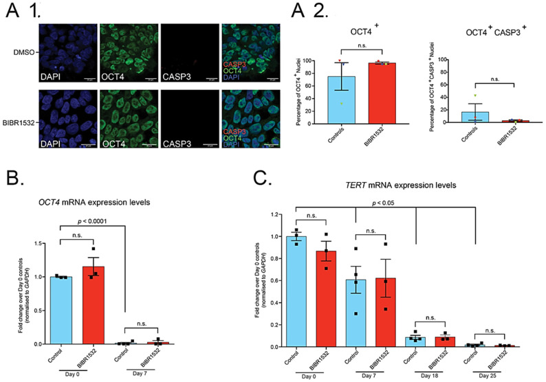
Several studies have shown that human induced pluripotent stem cell (iPSC)-derivatives are essentially fetal in terms of their maturational status. Inducing ageing in iPSC-motor neuron (MN) models of amyotrophic lateral sclerosis (ALS) has the potential to capture pathology with higher fidelity and consequently improve translational success. We show here that the telomerase inhibitor BIBR1532, hypothesised to recapitulate the telomere attrition hallmark of ageing in iPSC-MNs, was in fact cytotoxic to feeder-free iPSCs when used at doses previously shown to be effective in iPSCs grown on a layer of mouse embryonic fibroblasts. Toxicity in feeder-free cultures was not rescued by co-treatment with Rho Kinase (ROCK) inhibitor (Y-27632). Moreover, the highest concentration of BIBR1532 compatible with continued iPSC culture proved insufficient to induce detectable telomerase inhibition. Our data suggest that direct toxicity by BIBR1532 is the most likely cause of iPSC death observed, and that culture methods may influence enhanced toxicity. Therefore, recapitulation of ageing hallmarks in iPSC-MNs, which might reveal novel and relevant human disease targets in ALS, is not achievable in feeder-free culture through the use of this small molecule telomerase inhibitor.

Keywords: BIBR1532; TERT; ageing; amyotrophic lateral sclerosis (ALS); induced pluripotent stem cells (iPSC); motor neurons (MNs); telomerase.


Conflict of interest statement

The authors declare no conflict of interest.

Figures

Figure 1
Figure 1
Treatment of iPSCs with 0.05 µM BIBR1532 allows continued culture, maintains pluripotency, and does not induce apoptosis; however, 0.05 µM BIBR1532 has no effect on TERT expression throughout iPSC-MN directed differentiation. (A1) Qualitative immunocytochemistry for DAPI, OCT4, and activated CASP3 in iPSCs treated with 0.05 µM BIBR1532 or DMSO for 15 days. Scale bars = 20 µm. (A2) Quantitative analysis of the percentage of OCT4+ nuclei and OCT4+CASP3+ nuclei in 0.05 µM BIBR1532 treated iPSCs and DMSO controls. Data presented as mean ± S.E.M. Unpaired t-test. Three biological repeats (represented by datapoints), five fields per cover slip. (B) qRT-PCR analysis of OCT4 expression at day 0 and day 7 of neural induction, in cells treated with 0.05 µM BIBR1532 or DMSO, normalised over GAPDH expression, relative to day 0 control iPSCs. (C) qRT-PCR analysis of expression of TERT throughout iPSC-MN differentiation in cells treated with 0.05 µM BIBR1532 or DMSO, normalised to GAPDH expression, relative to day 0 control iPSCs. (B,C) Data presented as mean ± S.E.M. One experimental block, three biological replicates. Datapoints represent biological replicates. From day 7 onwards, an additional technical replicate of the Ctrl 1 line was included in controls, represented as an additional datapoint above. Two-way ANOVA, with Tukey’s test for multiple comparisons. n.s. = non-significant, with significance placed at a p-value of <0.05. p-values displayed graphically refer to the statistical difference between the indicated timepoint and day 0 control samples.

Similar articles

Cited by

References

    1. Ho R., Sances S., Gowing G., Amoroso M.W., O’Rourke J.G., Sahabian A., Wichterle H., Baloh R.H., Sareen D., Svendsen C.N. ALS disrupts spinal motor neuron maturation and aging pathways within gene co-expression networks. Nat. Neurosci. 2016;19:1256–1267. doi: 10.1038/nn.4345. - DOI - PMC - PubMed
    1. Herskovits A.Z., Hunter T.A., Maxwell N., Pereira K., Whittaker C.A., Valdez G., Guarente L.P. SIRT1 deacetylase in aging-induced neuromuscular degeneration and amyotrophic lateral sclerosis. Aging Cell. 2018;17:e12839. doi: 10.1111/acel.12839. - DOI - PMC - PubMed
    1. Das M.M., Svendsen C.N. Astrocytes show reduced support of motor neurons with aging that is accelerated in a rodent model of ALS. Neurobiol. Aging. 2015;36:1130–1139. doi: 10.1016/j.neurobiolaging.2014.09.020. - DOI - PubMed
    1. Kreiter N., Pal A., Lojewski X., Corcia P., Naujock M., Reinhardt P., Sterneckert J., Petri S., Wegner F., Storch A., et al. Age-dependent neurodegeneration and organelle transport deficiencies in mutant TDP43 patient-derived neurons are independent of TDP43 aggregation. Neurobiol. Dis. 2018;115:167–181. doi: 10.1016/j.nbd.2018.03.010. - DOI - PubMed
    1. Pandya V.A., Patani R. Decoding the relationship between ageing and amyotrophic lateral sclerosis: A cellular perspective. Brain. 2019;143:1057–1072. doi: 10.1093/brain/awz360. - DOI - PMC - PubMed

MeSH terms

LinkOut - more resources