Review
. 2021 Mar 23;22(6):3259.
doi: 10.3390/ijms22063259.

Structure and Function of Ion Channels Regulating Sperm Motility-An Overview

Affiliations
Review

Structure and Function of Ion Channels Regulating Sperm Motility-An Overview

Karolina Nowicka-Bauer et al. Int J Mol Sci. .

Abstract

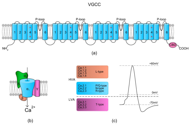
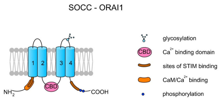
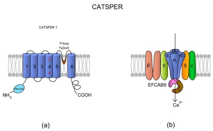
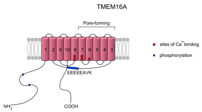
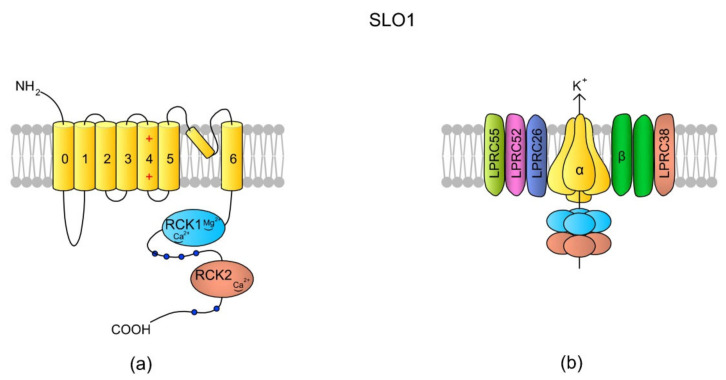
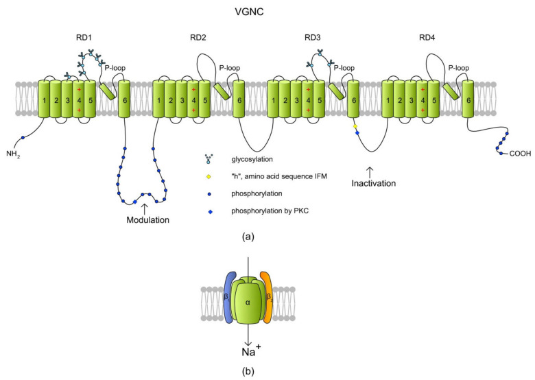
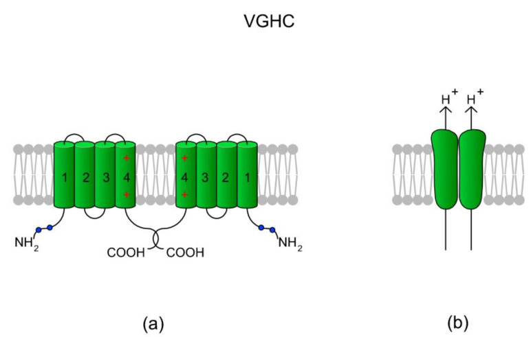
Sperm motility is linked to the activation of signaling pathways that trigger movement. These pathways are mainly dependent on Ca2+, which acts as a secondary messenger. The maintenance of adequate Ca2+ concentrations is possible thanks to proper concentrations of other ions, such as K+ and Na+, among others, that modulate plasma membrane potential and the intracellular pH. Like in every cell, ion homeostasis in spermatozoa is ensured by a vast spectrum of ion channels supported by the work of ion pumps and transporters. To achieve success in fertilization, sperm ion channels have to be sensitive to various external and internal factors. This sensitivity is provided by specific channel structures. In addition, novel sperm-specific channels or isoforms have been found with compositions that increase the chance of fertilization. Notably, the most significant sperm ion channel is the cation channel of sperm (CatSper), which is a sperm-specific Ca2+ channel required for the hyperactivation of sperm motility. The role of other ion channels in the spermatozoa, such as voltage-gated Ca2+ channels (VGCCs), Ca2+-activated Cl-channels (CaCCs), SLO K+ channels or voltage-gated H+ channels (VGHCs), is to ensure the activation and modulation of CatSper. As the activation of sperm motility differs among metazoa, different ion channels may participate; however, knowledge regarding these channels is still scarce. In the present review, the roles and structures of the most important known ion channels are described in regard to regulation of sperm motility in animals.

Keywords: calcium; chloride; ion channels; membrane channels; potassium; proton; sodium; sperm motility.


Conflict of interest statement

The authors declare no conflict of interest.

Figures

Figure 1
Figure 1
Voltage-gated Ca2+ channel (VGCC) structure scheme. (a) The topology of the α1 subunit is made up of four homologous domains that each consist of six transmembrane α helices (TM1–6). TM4 from each homologous domain serves as the voltage sensor moving outward and rotates under the influence of the electric field, thereby initiating a conformational change that opens the respective pore. TM5, TM6, and the loop between them (P-loop) from each domain form a pore. The C-terminal tail contains a Ca2+ binding domain (CBD) and in some types of VGCCs a site for calmodulin (calcium-modulated protein; CaM) binding. The binding of Ca2+ to CBD or via CaM inactivates the channels. (b) A schematic presentation of the VGCC subunits (α1, α2δ, β, and γ) with their spatial localizations. (c) Overview of the types of VGCCs in relation to Vm-dependent activation – high voltage activation (HVA) and low voltage activation (LVA) (based on References [26,27]).
Figure 2
Figure 2
Topology of a store-operated Ca2+ channel (SOCC) created by ORAI1. Each ORAI protein has four TMs. TM2 and TM3 create a pore. There are two sites for STIM1 binding at the N- and C-termini. The interaction between STIM1 and ORAI activates the channel and the release of Ca2+ from the endoplasmic reticulum (ER). The binding of Ca2+ by the Ca2+ binding domain (CBD) localized on the central loop inactivates the channel [51]. Additionally, it can also be inactivated upon CaM binding [52].
Figure 3
Figure 3
A topological and spatial structure of CatSper. (a) The α1 subunit created by CatSper1. Like most voltage-gated channels, each α subunit contains six transmembrane domains (TM1–TM6) creating two physiologically distinctive regions, namely the voltage-sensing domain (VSD; TM1–4) and pore-forming region (TM5–6). Each TM4 contains several (two to six) positively charged amino acid residues that serve as voltage sensors (reviewed in Reference [57]). Voltage slopes move TM4, resulting in conformational changes that open and close the channel pore [64]. Additionally, a short and hydrophobic cyclic structure linking TM5–6 contains a conserved homologous amino acid sequence (T × D × W), which selectively permits Ca2+ influx. The N-terminus of CatSper 1 contains a specific histidine-rich region that might be involved in the pH regulation of CatSper activity. (b) The topological localizations of all auxiliary subunits are not randomly organized. The auxiliary CatSperβ subunit has two predicted TMs that are separated by a large (ca. 1000 amino acids) extracellular loop [64], whereas CatSperγ, CatSperδ, and CatSperε feature only one TM. Brown et al. [69] suggested that CatSperζ is a late evolutionary adaptation to maximize fertilization success inside the female mammalian reproductive tract. The predicted topology of Hwang et al. [62] situates the CatSperζ and EFCAB9 subunits as a cytoplasm complex that is located just below the CatSper 1–4 subunits. This complex interacts with the channel pore as a gatekeeper. The increase in pHi causes Ca2+ binding to highly conserved EF-hands of EFCAB9, leading to dissociation of the EFCAB9-CatSperζ complex and full activation of the channel. Accordingly, EFCAB9-CatSperζ appears to be responsible for both modulation of the channel activity and organization of the CatSper domains [62]. The scheme has been prepared based on Reference [62].
Figure 4
Figure 4
A simplified topology of the TMEM16A monomer. Each monomer has 10 TMs. The ion conduction pore of TMEM16A is formed by TMs three to seven in each subunit, and thus the CaCC features two pores [96,97]. As summarized in a review of Ji et al. [97], the activation of TMEM16A is gated by two main mechanisms: voltage (Vm) and low concentrations of Ca2+ (<600 nM) via the EEEEEAVK motif in the TM2–TM3 loop. Contreras-Vite et al. [98] proposed a gating mechanism model where TMEM16A is directly activated by the Vm-dependent binding of two Ca2+ ions coupled by a Vm-dependent binding of one external Cl ion. The scheme was prepared based on Reference [97].
Figure 5
Figure 5
SLO1 structure scheme. (a) A topology of the α subunit. Each α subunit consists of seven (0–6) TMs, where TM4 is a typical voltage-sensing domain (VSD). An extracellular loop between TM5 and TM6 forms the pore. The N-tail is located extracellularly but the C-end is a long tail containing the RCK1 (regulator of K+ conductance 1) and RKC2 domains [135]. The structural difference between SLO1 and SLO3 is that there are “Ca2+-bowl” structures within the RKC domains of SLO1, making the channel sensitive to [Ca2+]i. (b) In the tetrameric structure of the channel, the cytoplasmic C-termini creates a gating ring. According to the literature, SLO1 has five auxiliary subunits: one β subunit (with two transmembrane domains) and four Leucine-rich repeat-containing membrane proteins (LRRCs, also named γ subunits), LRRC26, LRCC52, LRRC55, and LRRC38, which modulate SLO1 sensitivity to Vm and [Ca2+]i (revised by Reference [144]). In murine testes and spermatozoa, two auxiliary subunits of the SLO3 channel have been identified: Lrrc52 and Lrrc26. Both of them are involved in the regulation of SLO3, and the expression of Lrrc52 is critically dependent on the presence of SLO3 [143]. The schemes are adapted from References [136,144].
Figure 6
Figure 6
A structure of a voltage-gated Na+ channel (VGNC) based on a SCN2A isoform. (a) The α subunit is created by four repeat domains (RD1–RD4) that each have six TMs. Classically, TM1–TM4 of each domain form a VSD where TM4 acts as a positively charged sensor. During depolarization, TM4 is believed to move toward the extracellular surface, allowing the channel to become permeable to ions. Na+ is transported inside a cell through a pore (P-loop) formed between TM5 and TM6 of each RD. The RDs are connected with long intracytoplasmic loops with sites for protein phosphorylation via PKA and PKC [157]. The cytoplasmic loop between RD3 and RD4 contains an “h” (I × F × M sequence) motif, which stands for a hydrophobic triad of amino acids, namely, isoleucine, phenylalanine, and methionine (I1488, F1489, and M1490). The IFM motif is involved in the inactivation of VGNC, serving as a hydrophobic latch for a hinged lid formed by the loop between RD3 and RD4 [159]. Phosphorylation in the RD1/RD2 and RD3/RD4 loops modulates the channel inactivation (adapted from Reference [157], revised in Reference [160]). (b) A cartoon of VGNC created by the pore-forming α subunit and the two auxiliary β subunits.
Figure 7
Figure 7
A voltage-gated H+ channel (VGHC) and its structure. (a) A VGHC monomer is created by four TMs which in classical voltage-gated channels comprise VSD. Accordingly, VGHCs do not possess a pore-forming domain (TM5-TM6) and the extrusion of H+ ions probably takes place via a water wire spanning the VSD [168]. According to Boonamnaj et al. [169], in VGHC dimers, C-terminal tails interact by forming a coiled structure that stabilizes the channel. Sites of phosphorylation in the N-termini may enhance the selectivity of the channel. (b) A dimeric structure of a VGHC. As the VGHC has no pore-forming domains, H+ diffuses through each monomer.

References

    1. Watanabe A., Takayama-Watanabe E., Vines C.A., Cherr G.N. Sperm motility-initiating substance in newt egg-jelly induces differential initiation of sperm motility based on sperm intracellular calcium levels. Dev. Growth Differ. 2011;53:9–17. doi: 10.1111/j.1440-169X.2010.01216.x. - DOI - PubMed
    1. Takayama-Watanabe E., Ochiai H., Tanino S., Watanabe A. Contribution of different Ca2⁺ channels to the acrosome reaction-mediated initiation of sperm motility in the newt Cynops pyrrhogaster. Zygote. 2015;23:342–351. doi: 10.1017/S0967199413000609. - DOI - PubMed
    1. Hansbrough J.R., Garbers D.L. Speract. Purification and characterization of a peptide associated with eggs that activates spermatozoa. J. Biol. Chem. 1981;256:1447–1452. doi: 10.1016/S0021-9258(19)69983-7. - DOI - PubMed
    1. Ohtake H. Sperm-activating proteins obtained from the herring eggs. Fish Physiol. Biochem. 2003;28:199–202. doi: 10.1023/B:FISH.0000030527.87437.05. - DOI
    1. Alavi S.M.H., Cosson J., Bondarenko O., Linhart O. Sperm motility in fishes: (III) diversity of regulatory signals from membrane to the axoneme. Theriogenology. 2019;136:143–165. doi: 10.1016/j.theriogenology.2019.06.038. - DOI - PubMed

MeSH terms

LinkOut - more resources