. 2021 Mar 23;14(3):285.
doi: 10.3390/ph14030285.

The Influence of Oxidative Stress on Serum Albumin Structure as a Carrier of Selected Diazaphenothiazine with Potential Anticancer Activity

Affiliations

The Influence of Oxidative Stress on Serum Albumin Structure as a Carrier of Selected Diazaphenothiazine with Potential Anticancer Activity

Małgorzata Maciążek-Jurczyk et al. Pharmaceuticals (Basel). .

Abstract

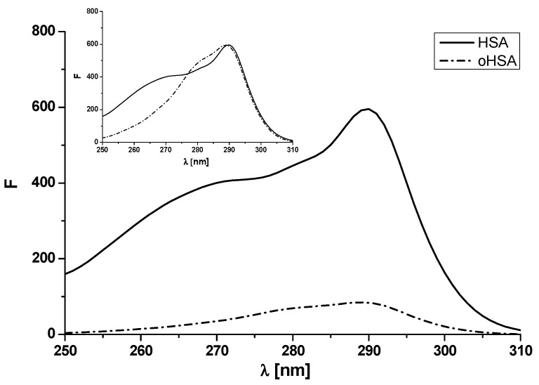
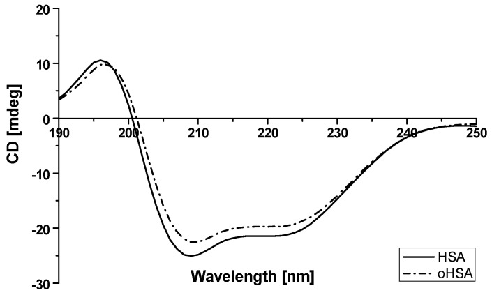
Albumin is one of the most important proteins in human blood. Among its multiple functions, drug binding is crucial in terms of drug distribution in human body. This protein undergoes many modifications that are certain to influence protein activity and affect its structure. One such reaction is albumin oxidation. Chloramine T is a strong oxidant. Solutions of human serum albumin, both non-modified and modified by chloramine T, were examined with the use of fluorescence, absorption and circular dichroism (CD) spectroscopy. 10H-3,6-diazaphenothiazine (DAPT) has anticancer activity and it has been studied for the first time in terms of binding with human serum albumin-its potential as a transporting protein. Using fluorescence spectroscopy, in the presence of dansylated amino acids, dansyl-l-glutamine (dGlu), dansyl-l-proline (dPro), DAPT binding with two main albumin sites-in subdomain IIA and IIIA-has been evaluated. Based on the conducted data, in order to measure the stability of DAPT complexes with human (HSA) and oxidized (oHSA) serum albumin, association constant (Ka) for ligand-HSA and ligand-oHSA complexes were calculated. It has been presumed that oxidation is not an important issue in terms of 10H-3,6-diazaphenothiazine binding to albumin. It means that the distribution of this substance is similar regardless of changes in albumin structure caused by oxidation, natural occurring in the organism.

Keywords: 10H-3,6-diazaphenothiazine; human serum albumin; oxidation; spectroscopic methods.


Conflict of interest statement

The authors declare no conflict of interest.

Figures

Figure 1
Figure 1
Structural formula of 10H-3,6-diazaphenothiazine (DAPT).
Figure 2
Figure 2
Absorption spectra of unmodified (human serum albumin (HSA)) and oxidized (oxidized human serum albumin (oHSA)) human serum albumin at 5 × 10⁻⁶ mol·L−1 concentration. In the insert second derivative of 5 × 10⁻⁶ mol·L−1 HSA and oHSA absorption spectrum.
Figure 3
Figure 3
Emission fluorescence spectra of unmodified (HSA) and oxidized (oHSA) human serum albumin at 5 × 10⁻⁶ mol·L−1 concentration excited at (a) λex 275 nm and (b) λex 295 nm.
Figure 4
Figure 4
Excitation fluorescence spectra of unmodified (HSA) and oxidized human serum albumin (oHSA) at 5 × 10⁻⁶ mol·L–1 concentration. In the insert normalized excitation fluorescence spectra of oHSA and unmodified HSA.
Figure 5
Figure 5
Circular dichroism (CD) spectra of HSA and oHSA at 1 × 10⁻⁶ mol·L–1 concentration.
Figure 6
Figure 6
Emission fluorescence spectra of (a) dGlu and (b) dPro in the presence of HSA and oHSA. [dGlu] = [dPro] 5 × 10⁻⁵ mol·L−1, [HSA] = [oHSA] 1 × 10⁻⁵ mol·L−1; λex = 350 nm.
Figure 7
Figure 7
Scatchard curve for (a) dGlu-HSA, dGlu-oHSA and (b) dPro-HSA, dPro-oHSA systems; λex 350 nm. [HSA] = [oHSA] 1 × 10⁻⁵ mol·L−1, [dGlu] 0.5 × 10⁻⁵ mol·L−1–6 × 10⁻⁵ mol·L−1. In the insert the binding isotherms for dPro-HSA and dPro-oHSA systems.
Figure 8
Figure 8
Comparison of quenching curves of (a) dGlu and (b) dPro at 5 × 10⁻⁶ mol·L−1 concentration in the presence of HSA, oHSA at 5 × 10⁻⁶ mol·L−1 concentration and DAPT (2.5 × 10⁻⁶ mol·L−1–8.5 × 10⁻⁵ mol·L−1); the error calculated as maximum deviation does not exceed the symbols.
Figure 9
Figure 9
The Stern-Volmer curves of F0/F vs. 10H-3,6-diazophenothiazine (DAPT) concentration at (a) λex 275 nm and (b) λex 295 nm; the error calculated as maximum deviation does not exceed the symbols.
Figure 10
Figure 10
The binding isotherms for DAPT-HSA and DAPT-oHSA systems at (a) λex 275 nm and (b) λex 295 nm.
Figure 11
Figure 11
The Klotz plots for DAPT-HSA and DAPT-oHSA systems at (a) λex 275 nm and (b) λex 295 nm.

Similar articles

Cited by

References

    1. Peters T. All about Albumin: Biochemistry, Genetics and Medical Applications. Academic Press; San Diego, CA, USA: 1995.
    1. Kragh-Hansen U., Chuang V.T.G., Otagiri M. Practical Aspects of the Ligand-Binding and Enzymatic Properties of Human Serum Albumin. Biol. Pharm. Bull. 2002;25:695–704. doi: 10.1248/bpb.25.695. - DOI - PubMed
    1. Spinella R., Sawhney R., Jalan R. Albumin in chronic liver disease: Structure, functions and therapeutic implications. Hepatol. Int. 2016;10:124–132. doi: 10.1007/s12072-015-9665-6. - DOI - PubMed
    1. Otagiri M., Chuang V.T.G. Albumin in Medicine: Pathological and Clinical Applications. Springer; Singapore: 2016.
    1. Fanali G., di Masi A., Trezza V., Marino M., Fasano M., Ascenzi P. Human serum albumin: From bench to bedside. Mol. Asp. Med. 2012;33:209–290. doi: 10.1016/j.mam.2011.12.002. - DOI - PubMed

LinkOut - more resources