Review
. 2021 Mar 23;13(6):981.
doi: 10.3390/polym13060981.

Recent Developments on Semiconducting Polymer Nanoparticles as Smart Photo-Therapeutic Agents for Cancer Treatments-A Review

Affiliations
Review

Recent Developments on Semiconducting Polymer Nanoparticles as Smart Photo-Therapeutic Agents for Cancer Treatments-A Review

N Sanoj Rejinold et al. Polymers (Basel). .

Abstract

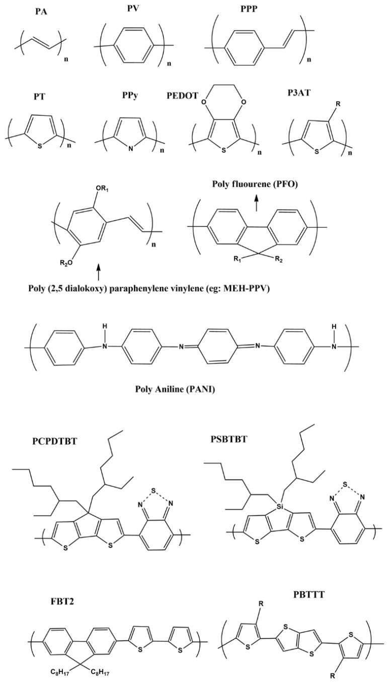
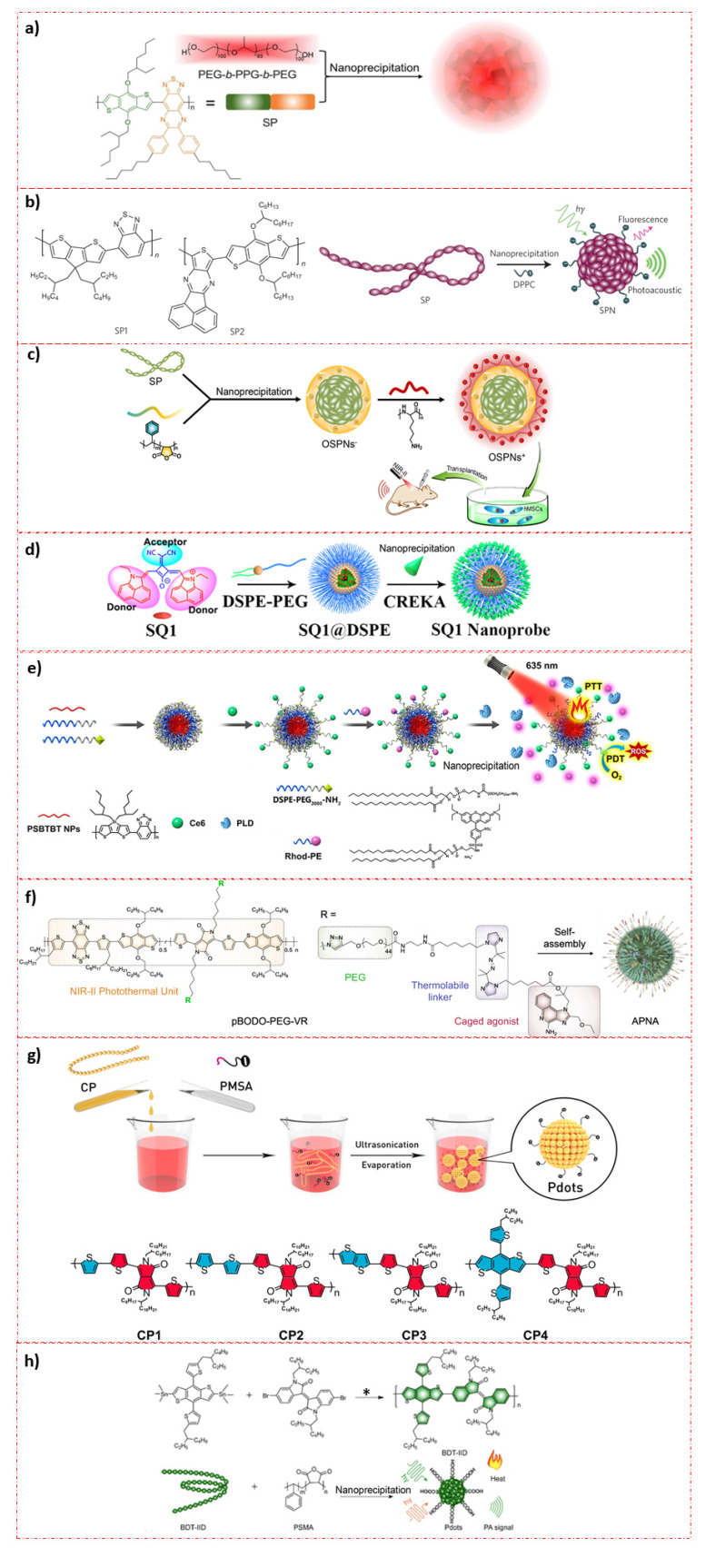
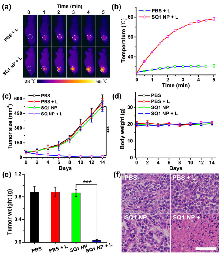
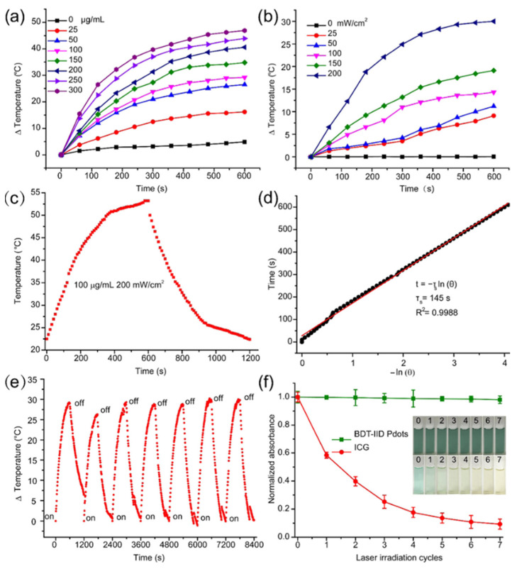
Semiconducting polymer nanoparticles (SPN) have been emerging as novel functional nano materials for phototherapy which includes PTT (photo-thermal therapy), PDT (photodynamic therapy), and their combination. Therefore, it is important to look into their recent developments and further explorations specifically in cancer treatment. Therefore, the present review describes novel semiconducting polymers at the nanoscale, along with their applications and limitations with a specific emphasis on future perspectives. Special focus is given on emerging and trending semiconducting polymeric nanoparticles in this review based on the research findings that have been published mostly within the last five years.

Keywords: applications; future scope; limitations; photo-therapy; semiconducting polymers.


Conflict of interest statement

The authors declare no conflict of interest. Additionally, the funders had no role in the design of the study; in the collection, analyses, or interpretation of data; in the writing of the manuscript, or in the decision to publish the results.

Figures

Figure 1
Figure 1
Shows various semi conducting polymers from first to third generations: PA-poly acetylene; PV-poly paraphenylene vinylene; PPP-poly para phenylene; PT-poly thiophene; PPy-poly pryrrole; PEDOT-poly ethylene dioxythiophene; P3AT-poly 3(alkyl) thiphene (R-Alkyl group); PCPDBT-poly(2,6-[4,4-bis-(2-ethylhexyl) 4H-cyclopenta (2,1-b;3,4-b1)dithiophene-alt-4,7-(2,1,3-benzothiadiazole)]; PSBTBT-Poly [(4,4′-bis(2-ethylhexyl)dithieno[3,2-b:2′,3′-d]silole)-2,6-diyl-alt-(2,1,3-benzothiadiazole)-4,6-diyl]; FBT2-poly(9,9-dioctylflourene-co-bithiophene); PBTTT-poly(2,5-bis(3-alkylthiophen-2-yl)thieno[3,2-b]thiophene respectively.
Figure 2
Figure 2
Preparative techniques for various SPN based photo-theranostic materials: (a) Illustration of the SPNs preparation from SP and PEG-b-PPG-b-PEG using nanoprecipitation method. (Reprinted Permission from American Chemical Society, 2020) [46]; (b) Molecular structures of SP1 and SP2 used for the preparation of SPN1 and SPN2, respectively. SPNs made through nanoprecipitation. SP is represented as a long chain of chromophore units (red oval beads). DPPC contains a short hydrophobic tail and a charged head and is illustrated as a string with a dark green ball at its end. (Reprinted permission from Nature Nanotechnology, 2014) [69]; (c) illustration of the preparation procedure of OSPNs+ and the photoacoustic labeling of hMSCs after transplantation. (Reprinted permission from American Chemical Society, 2018) [70]; (d) Molecular engineering and nano functionalization of Squaraine dye SQ1 for NIR-II/PA Bimodal Imaging and Photo-thermal ablation of metastatic breast cancer. (Reprinted permission from American Chemical Society, 2020) [74]; (e) Schematic Illustration of PLD-Activatable Tumor Image and PTT/PDT Combined Therapy (Reprinted permission from American Chemical Society, 2021) [75]; (f) Chemical structure of pBODO-PEG-VR and preparation of APNA (Reprinted permission from Nature, 2021) [76]; (g) Schematic illustration of preparation for Pdots (Reprinted permission from American Chemical Society, 2016) [48]; (h) Synthetic route of conjugated polymer BDT-IID (*) Pd(PPh3)4 and toluene, 110 °C and preparation of BDT-IID Pdots for PAI-guided PTT (Reprinted permission from American Chemical Society, 2018) [49].
Figure 3
Figure 3
PTT effects of Squarine dye based copolymer NPs on breast tumor burden on s mice. (a) Thermal imaging; (b) Thermal variations in tumor during PTT; (c) Tumor growth inhibition and (d) body weight analysis during treatment period; (e) Weight measures of tumor lesions post treatment with PBS, PBS + L, SQ1 NP, SQ1 NP + L. L refers NIR laser (915 nm, 0.5 W/cm2). Data present as mean ± SD, n = 5 (*** p < 0.01). (f) Histochemical analysis of tumor sections treated with PBS, PBS + L, SQ1 NP, SQ1 NP + L. The scale bar is 50 μm. (Reprinted permission American Chemical Society, 2020) [74].
Figure 4
Figure 4
PTT characteristics of BDT-IID Pdots. PTT heating curves of the BDT-IID Pdots (a) with different concentrations upon 200 mW/cm2 laser exposure at 660 nm and (b) with different laser power densities at 100 μg/mL. (c) Photothermal effect of the BDT-IID Pdots dispersions under laser irradiation at 660 nm (200 mW/cm2). Irradiation terminated after 600 s. (d) Time constant for heat transfer was determined to be τs = 145 s by applying the linear time data from the cooling period (after 600 s) versus negative natural logarithm of driving force temperature, obtained from the cooling stage of (c); (e) thermal variations of the Pdots under laser exposure at 200 mW/cm2 for seven light on/off cycles (10 min of irradiation for each cycle). (f) Change in absorbance intensity of BDT-IID Pdots and ICG after repeated laser irradiation (n = 7). The figure inserts show the changes of BDT-IID Pdots and ICG after repeated laser irradiation (n = 7). (Reprinted Permission American Chemical Society 2018) [49].
Figure 5
Figure 5
Real-time in-vivo NIR-II fluorescence microscopic imaging of mouse brain vasculature. (a) Cerebrovascular imaging at various depths (100–900 μm) after the intravenous injection of L1057 NPs. The excitation wavelength was 980 nm. Scale bar: 100 μm. (b) Cross-sectional fluorescence intensity profiles (and Gaussian fits (red) with fwhm indicated by arrows) along the red lines circled with green in panel a; PTT efficacy of L1057 NPs on tumors. (c,d) PTT images (c) and corresponding temperature changes (d) of 4T1-tumor-bearing mice under irradiation with an 808 (0.33 W/cm2) or 980 nm (0.72 W/cm2) laser. (e,f) Body weight (e) and tumor volume (f) curves of tumor-bearing mice at different time points after receiving PTT. (g,h) Tumor weight (g) and H&E staining (h) of the tumor tissues from mice sacrificed at day 18 post-PTT treatment. Scale bar: 100 μm. Results are presented as the mean ± S.D., n = 5. Statistical significance was calculated using one-way ANOVA with the Tukey posthoc test. *** p < 0.001. (Reprinted Permission from American Chemical Society, 2020) [61].
Figure 6
Figure 6
In-vivo photodynamic effect of Ce6-doped Pdots. (a) representative photographs for tumor-bearing nude mice during PDT. Relative tumor volume (V/V0) (b) and body weight (c) of tumor-bearing nude mice in control group (Con), intravenous injection group (I.V.), intratumoral injection low-dose group (I.T. low-dose) and intratumoral injection high dose group (I.T. high-dose). (d) Histopathology analysis of tumors and organs collected from tumor bearing nude mice after PDT. (Reprinted Permission American Chemical Society, 2017) [91].
Figure 7
Figure 7
Schematic illustrations of the preparation of NPs for a PAI-Guided PDT/PTT and proposed photo-physical mechanism (Reprinted Permission American Chemical Society, 2019) [104].
Figure 8
Figure 8
(a) Chemical structure of pBODO-PEG-VR and preparation of APNA. (b) Mechanism of antitumor immune response by APNA-mediated NIR-II photothermal immunotherapy. TAAs tumor-associated antigens, DAMPs damage-associated molecular patterns, iDC immatureDC, mDC mature DC, HMGB1 high-mobility group box 1 protein (Reprinted Permission from Nature, 2021 under creative common license) [76].
Figure 9
Figure 9
(a) Synthetic procedures. First, sub-50 nm SP brush/fluorocarbon/phenylene triple-hybridized HPFON prepared by deposition of bissilylated organosilica precursors onto an MSN template via hydrolysis based on the chemical homology principle and selective MSN etching through an ammonia-assisted hot water etching strategy. Then, an in situ polymerization method is applied to conjugate alkyl chains and PEG polymers onto the inner and outer shell of the HPFON for enhanced hydrophobic drug loading as well as improved biocompatibility. Finally, SNAP and O2 were loaded onto the resultant pHPFON to generate the pHPFON-NO/O2. (b) Schematic illustration of the binary “reducing expenditure and broadening sources” tumor oxygenation strategy by programable delivery of NO and O2 with pHPFON-NO/O2 to overcome hypoxia-associated therapy resistance for boosted anti-cancer radiotherapy. (Reprinted Permission from Nature, 2021 under creative common license) [126].

References

    1. Song M. Cancer overtakes vascular disease as leading cause of excess death associated with diabetes. Lancet Diabetes Endocrinol. 2021;9:131–133. doi: 10.1016/S2213-8587(21)00016-4. - DOI - PubMed
    1. Pinheiro P.S., Callahan K.E., Jones P.D., Morris C., Ransdell J.M., Kwon D., Brown C.P., Kobetz E.N. Liver cancer: A leading cause of cancer death in the United States and the role of the 1945–1965 birth cohort by ethnicity. JHEP Rep. 2019;1:162–169. doi: 10.1016/j.jhepr.2019.05.008. - DOI - PMC - PubMed
    1. Demarinis S. Cancer overtakes cardiovascular disease as leading cause of death in wealthy nations. Explore. 2020;16:6–7. doi: 10.1016/j.explore.2019.11.003. - DOI - PubMed
    1. Mahase E. Cancer overtakes CVD to become leading cause of death in high income countries. BMJ. 2019;366:l5368. doi: 10.1136/bmj.l5368. - DOI - PubMed
    1. Hastings K.G., Kapphahn K., Boothroyd D.B., Rehkopf D.H., Cullen M.R., Palaniappan L. Transition From Heart Disease to Cancer as the Leading Cause of Death in the United States. Ann. Intern. Med. 2019;171:225. doi: 10.7326/L19-0203. - DOI - PubMed

LinkOut - more resources