Review
. 2021 Mar 23;9(3):33.
doi: 10.3390/dj9030033.

Sport and Dental Traumatology: Surgical Solutions and Prevention

Affiliations
Review

Sport and Dental Traumatology: Surgical Solutions and Prevention

Lorenzo Mordini et al. Dent J (Basel). .

Abstract

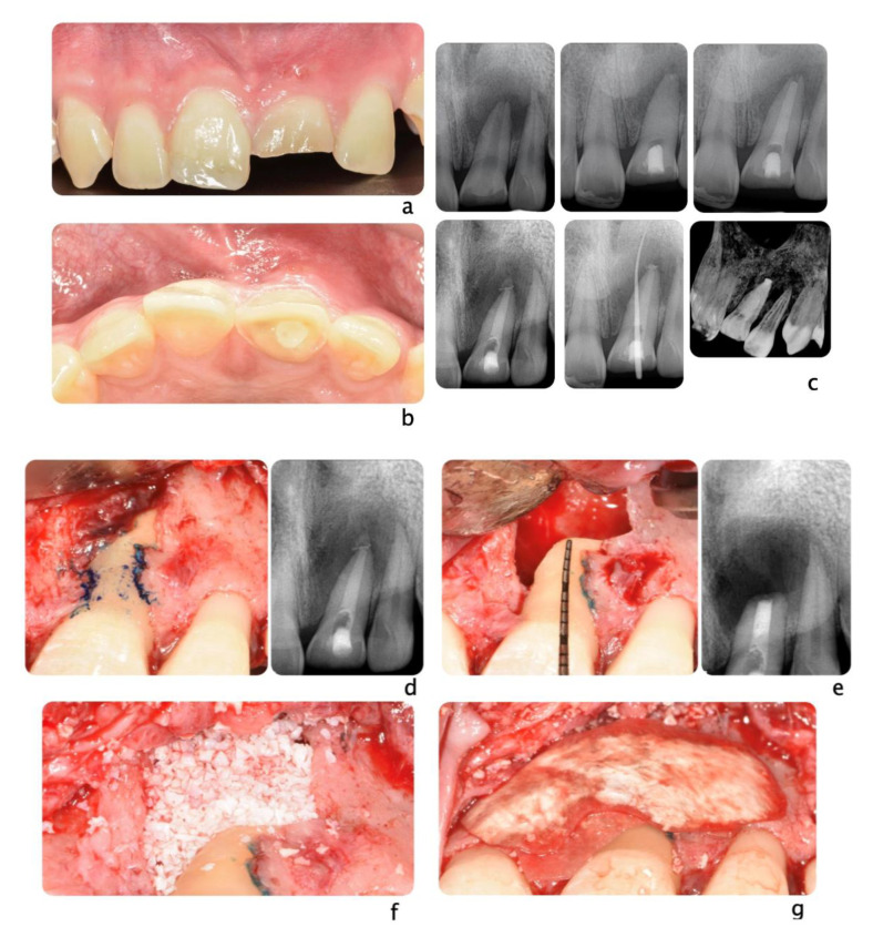
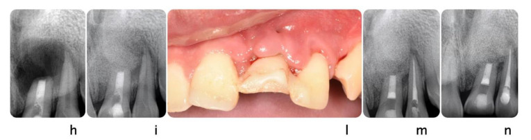
Trauma is a worldwide cause of millions of deaths and severe injuries every year, all over the world. Despite the limited extension of the oral region compared to the whole body, dental and oral injuries account for a fairly high percentage of all body traumas. Among head and neck traumas, dental and facial injuries are highly correlated to sport activities, and their management can be a real challenge for practitioners of any specialty. In case of trauma directed to periodontal structures, restorative and endodontic solutions may not be sufficient to achieve a definitive and long-lasting treatment. This article aims to illustrate surgical options and appliances to prevent dental injuries that may be available to the clinicians treating dental trauma involving oral soft and hard tissues.

Keywords: dental implants; dental trauma; facial injury; periodontology; tooth auto-transplantation.


Conflict of interest statement

Authors report no conflict of interest.

Figures

Figure 1
Figure 1
This radiograph shows tooth#24 horizontal fracture and radiolucency in the anterior mandible, as a result of a blow received from the patient playing boxing.
Figure 2
Figure 2
Ski accident that involved a 26-year-old male. He was treated in the emergency room with a metal retainer anchored with orthodontic wire around teeth involved in the trauma (a). The diagnosis was non-complicated maxillary fracture and teeth #7, 8, and 10 concussion and #9 avulsion. Panoramic image of the metal retainer (b). After 1 month of healing, metal retainer was removed (c) and teeth #7, 8, and 10 diagnosed as necrotic. Root canal treatments were performed (d).
Figure 3
Figure 3
A 20-year-old Asian male had #8 diagnosed with a root fractured due to sport related trauma (a). The tooth was endodontically treated, followed by healing with interposition of connective tissue (b). After healing was completed, a second sport injury involved the same tooth. The tooth mobility increased, and a periodontal lesion was diagnosed by elevated probing depth. The tooth was stabilized with orthodontic wire and patient was referred to periodontist for evaluation. Combined with malocclusion and anterior open-bite, the treatment plan was made as full-month orthodontics and auto-transplantation of #28. Tooth #8 and 28 were extracted (c,d) and a premolar replica was printed (d). After socket adjustment with the replica (e), tooth #28 was stabilized in place with sutures (f). After periodontal stabilization and verification of periodontal healing (g), the final restoration was delivered (h).
Figure 4
Figure 4
A 24-year-old female fell from her bike during a race. She hit the tarmac and resulted in losing teeth#9, 10 and 11 (a,b) as well as a portion of the alveolar bone (c) as seen on the 3D print of the maxilla. An incisal chip on tooth #8 completed the damage of the fall. After an analysis of residual hard and soft tissue volumes, a digital wax-up was created to plan the future implant placement and restorations (d). Guided tissue regeneration was performed, and implants were placed in a Type 4 timeline (e,f). A provisional fixed partial denture and connective tissue graft were inserted to improve esthetics and tissue conditioning (g).
Figure 5
Figure 5
Clinical scenario of a 17 years-old adolescent hit by a baseball ball in the anterior maxillary region. The boy presented to the Periodontal Department at Tufts University, Boston USA with crown fracture of left central incisor (#9) (a,b). Peri-apical radiograph show apical radiolucency, sign of necrosis. After the diagnosis, CaOH2 was applied. The root canal definitive treatment was completed but after 2 months the patient still presented with a fistula, that was tracked via a gutta-percha point. A CBCT scan was performed in order to diagnose the extent of the peri-apical lesion (c in sequence). The extent of the lesion did not suggest an endodontic therapy revision. Exploratory surgery was performed in order to rule out tooth fracture (d). The apex was resected in order to access the palatal aspect of the tooth. A PA radiograph was taken in order to verify correct apex resection and endodontic retrograde seal (e). Due to active patient skeletal growth, a decision was made to enucleate the endodontic cyst and treat the cavity with bone grafting material, in order to preserve the site for future implant placement (f,g). PA radiograph comparison before and after grafting placement (h,i). The patient was followed up for 2 months, and a fistula was identified apical to #9 (l). Tooth #10 was diagnosed as necrotic. A root canal was performed (m) and the apical radiolucency and fistula were resolved at 1 month follow up (n).
Figure 5
Figure 5
Clinical scenario of a 17 years-old adolescent hit by a baseball ball in the anterior maxillary region. The boy presented to the Periodontal Department at Tufts University, Boston USA with crown fracture of left central incisor (#9) (a,b). Peri-apical radiograph show apical radiolucency, sign of necrosis. After the diagnosis, CaOH2 was applied. The root canal definitive treatment was completed but after 2 months the patient still presented with a fistula, that was tracked via a gutta-percha point. A CBCT scan was performed in order to diagnose the extent of the peri-apical lesion (c in sequence). The extent of the lesion did not suggest an endodontic therapy revision. Exploratory surgery was performed in order to rule out tooth fracture (d). The apex was resected in order to access the palatal aspect of the tooth. A PA radiograph was taken in order to verify correct apex resection and endodontic retrograde seal (e). Due to active patient skeletal growth, a decision was made to enucleate the endodontic cyst and treat the cavity with bone grafting material, in order to preserve the site for future implant placement (f,g). PA radiograph comparison before and after grafting placement (h,i). The patient was followed up for 2 months, and a fistula was identified apical to #9 (l). Tooth #10 was diagnosed as necrotic. A root canal was performed (m) and the apical radiolucency and fistula were resolved at 1 month follow up (n).

References

    1. World Health Organization . Injuries and Violence: The Facts 2014. World Health Organization; Geneva, Switzerland: 2014.
    1. Petersson E.E., Andersson L., Sörensen S. Traumatic Oral Vs Non-Oral Injuries. Swed. Dent. J. 1997;21:55–68. - PubMed
    1. Hayter J.P., Ward A.J., Smith E.J. Maxillofacial Trauma in Severely Injured Patients. Br. J. Oral Maxillofac. Surg. 1991;29:370–373. doi: 10.1016/0266-4356(91)90003-N. - DOI - PubMed
    1. Piccininni P., Clough A., Padilla R., Piccininni G. Dental and Orofacial Injuries. Clin. Sports Med. 2017;36:369–405. doi: 10.1016/j.csm.2016.12.001. - DOI - PubMed
    1. Borssén E., Källestål C., Holm A.-K. Treatment time of traumatic dental injuries in a cohort of 16-year-olds in northern Sweden. Acta Odontologica Scandinavica. 2002;60:265–270. doi: 10.1080/00016350260248229. - DOI - PubMed

LinkOut - more resources