Review
. 2021 Mar 23;22(6):3276.
doi: 10.3390/ijms22063276.

New Insights into the Mammalian Egg Zona Pellucida

Affiliations
Review

New Insights into the Mammalian Egg Zona Pellucida

Carla Moros-Nicolás et al. Int J Mol Sci. .

Abstract

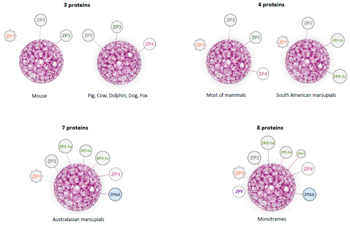
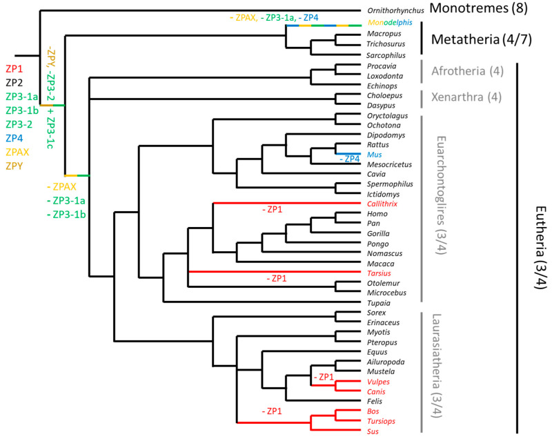
Mammalian oocytes are surrounded by an extracellular coat called the zona pellucida (ZP), which, from an evolutionary point of view, is the most ancient of the coats that envelope vertebrate oocytes and conceptuses. This matrix separates the oocyte from cumulus cells and is responsible for species-specific recognition between gametes, preventing polyspermy and protecting the preimplantation embryo. The ZP is a dynamic structure that shows different properties before and after fertilization. Until very recently, mammalian ZP was believed to be composed of only three glycoproteins, ZP1, ZP2 and ZP3, as first described in mouse. However, studies have revealed that this composition is not necessarily applicable to other mammals. Such differences can be explained by an analysis of the molecular evolution of the ZP gene family, during which ZP genes have suffered pseudogenization and duplication events that have resulted in differing models of ZP protein composition. The many discoveries made in recent years related to ZP composition and evolution suggest that a compilation would be useful. Moreover, this review analyses ZP biosynthesis, the role of each ZP protein in different mammalian species and how these proteins may interact among themselves and with other proteins present in the oviductal lumen.

Keywords: ZP; composition; mammals; molecular evolution; pseudogenization; zona pellucida.


Conflict of interest statement

The authors declare no conflict of interest.

Figures

Figure 1
Figure 1
The zona pellucida (ZP) of rabbit (A) and bovine (C) oocytes captured by conventional Hoffmann inverted microscopy appears very similar. The same zona pellucida captured by polarized light microscopy (B,D) reveals a different multilaminar structure and distinctly birefringence, and the meiotic spindle is easily appreciated (arrowhead). Scale bars 50 µm.
Figure 2
Figure 2
Zona pellucida composition in mammals. The ZP is composed of three, four, seven, or eight proteins. In house mouse (Mus musculus), the ZP is composed by ZP1, ZP2, and ZP3. In pig, cow, dolphin, dog, and fox, the ZP is formed by ZP2, ZP3, and ZP4. In most of mammals, the ZP is formed by four proteins: ZP1, ZP2, ZP3, and ZP4. In marsupials, two scenarios are found: four proteins in South American marsupials (ZP1, ZP2, ZP3-1b, and ZP3-1c) or seven proteins in Australasian marsupials (ZP1, ZP2, ZP3-1a, ZP3-1b, ZP3-1c, ZP4, ZPAX). In monotremes, the ZP is composed of eight ZP proteins: ZPY, ZP1 ZP2, ZP3-1a, ZP3-1b, ZP3-2, ZP4, ZPAX. In eutherian mammals ZP3-1c is written as ZP3 in order not to complicate the current nomenclature.
Figure 3
Figure 3
Evolution of ZP genes in mammals. Six ZP genes were probably present in the common ancestor. The gain and the loss of a gene in the phylogeny are indicated respectively by a plus or minus sign with the name of the gene concerned.
Figure 4
Figure 4
Ultrastructural analysis of the endoplasmic reticulum in the mice oocytes during the folliculogenesis. (A) Bilaminar primary ovarian follicle, semithin sections (right panel) and transmission electron microscope (panel left) immunostaining with anti-PDI antibody. The circular structure was specifically labelled with the anti-PDI antibody. (B) Multilaminar primary ovarian follicle, semithin sections (right panel), and transmission electron microscope (left panel) immunostaining with anti-PDI antibody. Note the presence of small dark vesicles (arrows) distributed throughout ooplasm specifically labelled with ER marker (Jiménez-Movilla M, Avilés M, Castells MT, Ballesta. Ultrastructural analysis of the endoplasmic reticulum in the mice oocytes during the folliculogenesis. First International Congress of Histology and Tissue Engineering, Spain, 2005).
Figure 5
Figure 5
Electron microscopical analysis of oocyte organelles involved in protein trafficking. (A) Human prophase I oocyte immunolabeled with anti-human ZP3 antibody. The Golgi apparatus showed a moderate reactivity. (B) Inmatured mice oocytes. Multivesicular aggregates (MVA) consisted of multiple vesicles embedded in an amorphous material, and the majority of them were found in close proximity to the oolemma. (C) Human prophase I oocyte immunolabeled with anti-human ZP3 antibody. Lysosomes like structures were strongly reactive (arrows) M: Mitochondria.

Similar articles

Cited by

References

    1. Modliński J.A. The role of the zona pellucida in the development of mouse eggs in vivo. J. Embryol. Exp. Morphol. 1970;23:539–547. - PubMed
    1. Bleil J.D., Wassarman P.M. Mammalian sperm-egg interaction: Identification of a glycoprotein in mouse egg zonae pellucidae possessing receptor activity for sperm. Cell. 1980;20:873–882. doi: 10.1016/0092-8674(80)90334-7. - DOI - PubMed
    1. Fahrenkamp E., Algarra B., Jovine L. Mammalian egg coat modifications and the block to polyspermy. Mol. Reprod. Dev. 2020;87:326–340. doi: 10.1002/mrd.23320. - DOI - PMC - PubMed
    1. Florman H.M., Storey B.T. Mouse gamete interactions: The zona pellucida is the site of the acrosome reaction leading to fertilization in vitro. Dev. Biol. 1982;91:121–130. doi: 10.1016/0012-1606(82)90015-X. - DOI - PubMed
    1. Berger T., Turner K.O., Meizel S., Hedrick J.L. Zona pellucida-induced acrosome reaction in boar sperm. Biol. Reprod. 1989;40:525–530. doi: 10.1095/biolreprod40.3.525. - DOI - PubMed

LinkOut - more resources