Review
. 2021 Mar 31;22(7):3613.
doi: 10.3390/ijms22073613.

A Comprehensive Review of Genetically Engineered Mouse Models for Prader-Willi Syndrome Research

Affiliations
Review

A Comprehensive Review of Genetically Engineered Mouse Models for Prader-Willi Syndrome Research

Delf-Magnus Kummerfeld et al. Int J Mol Sci. .

Abstract

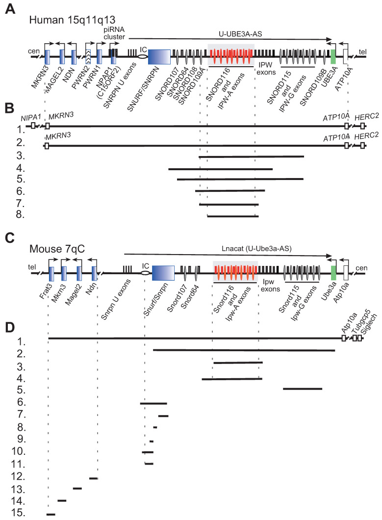
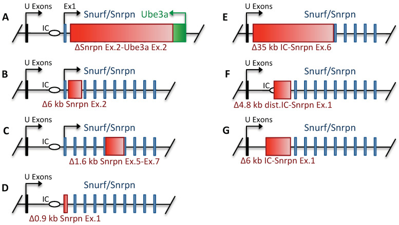
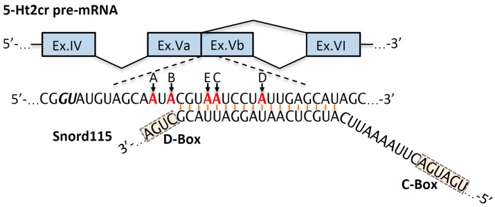
Prader-Willi syndrome (PWS) is a neurogenetic multifactorial disorder caused by the deletion or inactivation of paternally imprinted genes on human chromosome 15q11-q13. The affected homologous locus is on mouse chromosome 7C. The positional conservation and organization of genes including the imprinting pattern between mice and men implies similar physiological functions of this locus. Therefore, considerable efforts to recreate the pathogenesis of PWS have been accomplished in mouse models. We provide a summary of different mouse models that were generated for the analysis of PWS and discuss their impact on our current understanding of corresponding genes, their putative functions and the pathogenesis of PWS. Murine models of PWS unveiled the contribution of each affected gene to this multi-facetted disease, and also enabled the establishment of the minimal critical genomic region (PWScr) responsible for core symptoms, highlighting the importance of non-protein coding genes in the PWS locus. Although the underlying disease-causing mechanisms of PWS remain widely unresolved and existing mouse models do not fully capture the entire spectrum of the human PWS disorder, continuous improvements of genetically engineered mouse models have proven to be very powerful and valuable tools in PWS research.

Keywords: Magel2; PWS imprinting center (IC); Prader-Willi syndrome (PWS); Snord116; mouse models; non-coding RNAs.


Conflict of interest statement

The authors declare no conflict of interest.

Figures

Figure 1
Figure 1
Organization of human and mouse PWS loci, deletions in human and PWS mouse models are indicated. (A) Schematic representation of the human PWS locus on chromosome 15q11-q13. Blue rectangles denote paternally imprinted protein coding genes. Thin ovals show snoRNA gene locations; the imprinting center (IC) is denoted by a horizontal oval. Thin rectangles above the midline depict non-protein coding exons. SNORD116 and IPW-A exons are displayed in red and further highlighted by a grey rectangle. Arrows indicate promoters and the direction of transcription. The long arrow on top shows the putative U-UBE3A antisense transcript harboring the SNORD116 and SNORD115 clusters. Centromere and telomere regions are indicated as cen and tel. (B) Schematic representation of PWS chromosomal deletions. Lines 1 and 2 indicate the common 5–6 Mb PWS deletion [2]. Lines 38 represent the characterized PWS cases with microdeletion in the Snord116 array. Line 3.—[34], 4.—[35], 5.—[36], 6.—[37], 7.—[38], 8.—[39] (C) Schematic representation of the mouse PWS-locus on chromosome 7qC (symbols as above). (D) Schematic representation of available mouse models in PWS research. (1.) The largest chromosomal deletion that eliminates the PWS/AS region and a large portion of non-imprinted genes [40]. (2.) Deletion of the mouse PWS-locus span from the Snurf/Snrpn to Ube3a genes [41]. (3.) Deletion of the PWS critical region (~300 kb) (PWScr) comprising of Snord116 and IPW-A gene arrays [42]. (4.) The Snord116del mouse model eliminating a larger ~350 kb PWScr genomic region [43]. Note, that the genomic assembly of PWScr is not completed, a gap of ~50 kb inside the Snord116 cluster might increase the snoRNA gene copy number and overall size of deletion (UCSC, GRCm39/mm39 chr7:59457067- 59507068). (5.) Deletion of the Snord115 gene cluster [44] (6.11.) Deletions within the Snurf/Snrpn and PWS IC center (Details in Figure 2). (6.) 35 kb deletion of IC center and Snurf/Snrpn exons 1–6 [45]. (7.) Deletion of Snurf/Snrpn exon 6 including parts of exons 5 and 7 [45]. (8.) Deletion of Snurf/Snprn exon 2 [41]. (9.11.) Elimination of Snurf/Snrpn exon 1 and upstream genomic region: 0.9 kb (8), 4.8 kb (9) and 6 kb (10) deletions, respectively [46,47]. (12.–15.) Deletion of protein coding genes within the PWS-locus: (12.) Ndn [48,49,50,51]; (13.) Magel2 [52,53]; (14.) Mkrn3 [54]; (15.) Frat3 [30].
Figure 2
Figure 2
Schematic representation of genetically engineered mouse models harboring deletions within Snurf/Snrpn and PWS IC center (drawings are not to scale). Green and thin blue rectangles denote Ube3a and Snurf/Snrpn CDS exons, respectively. The red rectangles indicate generated genomic deletions. Thin black rectangles show location of U-exons; the imprinting center (IC) is denoted by a horizontal oval. (A). Large deletion of the genomic region between Snurf/Snrpn exon 2 and Ube3a exon 2. [41]. (B). Inactivation of the Snurf ORF by deleting the 6 kb region including Snurf/Snrpn exon 2 [41]. (C). Intragenic deletion of 1.6 kb within Snurf/Snrpn including exon 6 and parts of exons 5 and 7 disrupting the Snrpn ORF [45]. (D). Small 0.9 kb deletion of the major Snurf/Snrpn promoter together with first CDS exon [46]. (E). The PWS IC deletion, spanning 35 kb (originally described as 42 kb) including Snurf/Snrpn exons 1–6 [45]. (F). Deletion of 4.8 kb (later revealed to be 5.07 kb in size) genomic region, including Snurf/Snrpn exon 1 and the distal part of the PWS IC [46]. (G). The 6 kb deletion comprising PWS IC and Snurf/Snrpn exon 1 [47].
Figure 3
Figure 3
Schematic representation of 5-Ht2cr pre-mRNA exons IV to VI and putative targeting region of Snord115 within exon Vb (drawings are not to scale). Alternative splicing site (GU) of 5-Ht2cr pre-mRNA exon Vb is indicated with bold italic letters. A-to-I editing sites A–D are denoted in red and labeled with arrows, accordingly. Snord115 C- and D-boxes are highlighted [128,139].

Similar articles

Cited by

References

    1. McCandless S.E. Clinical report—Health supervision for children with Prader-Willi syndrome. Pediatrics. 2011;127:195–204. doi: 10.1542/peds.2010-2820. - DOI - PubMed
    1. Cassidy S.B., Schwartz S., Miller J.L., Driscoll D.J. Prader-Willi syndrome. Genet. Med. 2012;14:10–26. doi: 10.1038/gim.0b013e31822bead0. - DOI - PubMed
    1. Kalsner L., Chamberlain S.J. Prader-Willi, Angelman, and 15q11-q13 Duplication Syndromes. Pediatr. Clin. N. Am. 2015;62:587–606. doi: 10.1016/j.pcl.2015.03.004. - DOI - PMC - PubMed
    1. Angulo M.A., Butler M.G., Cataletto M.E. Prader-Willi syndrome: A review of clinical, genetic, and endocrine findings. J. Endocrinol. Invest. 2015;38:1249–1263. doi: 10.1007/s40618-015-0312-9. - DOI - PMC - PubMed
    1. Butler M.G., Miller J.L., Forster J.L. Prader-Willi Syndrome—Clinical Genetics, Diagnosis and Treatment Approaches: An Update. Curr. Pediatr. Rev. 2019;15:207–244. doi: 10.2174/1573396315666190716120925. - DOI - PMC - PubMed

Substances

LinkOut - more resources