Review
. 2021 Mar 31;10(7):1392.
doi: 10.3390/jcm10071392.

Assessment of Gastrointestinal Autonomic Dysfunction: Present and Future Perspectives

Affiliations
Review

Assessment of Gastrointestinal Autonomic Dysfunction: Present and Future Perspectives

Ditte S Kornum et al. J Clin Med. .

Abstract

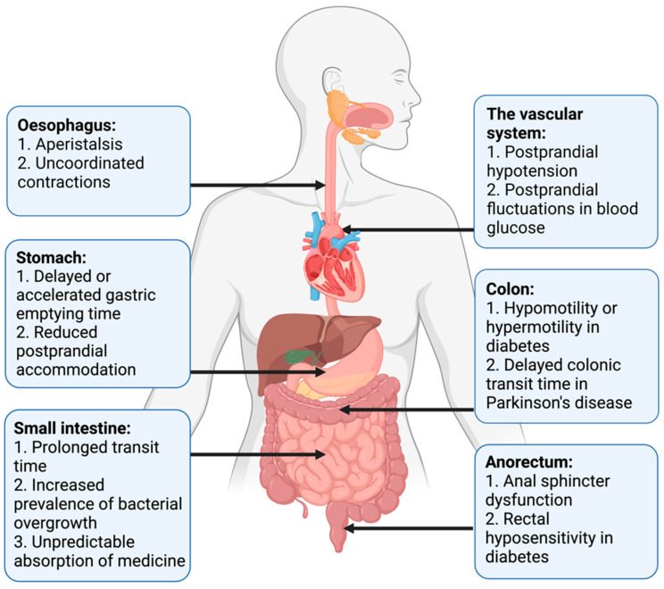
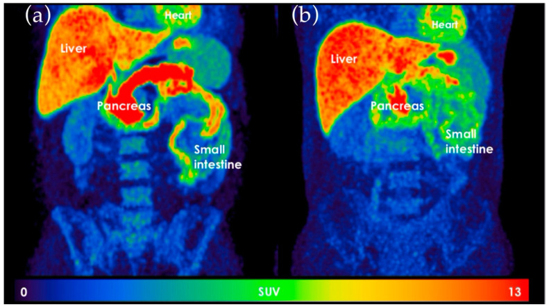
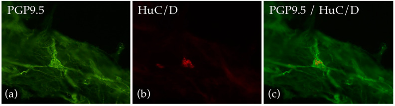
The autonomic nervous system delicately regulates the function of several target organs, including the gastrointestinal tract. Thus, nerve lesions or other nerve pathologies may cause autonomic dysfunction (AD). Some of the most common causes of AD are diabetes mellitus and α-synucleinopathies such as Parkinson's disease. Widespread dysmotility throughout the gastrointestinal tract is a common finding in AD, but no commercially available method exists for direct verification of enteric dysfunction. Thus, assessing segmental enteric physiological function is recommended to aid diagnostics and guide treatment. Several established assessment methods exist, but disadvantages such as lack of standardization, exposure to radiation, advanced data interpretation, or high cost, limit their utility. Emerging methods, including high-resolution colonic manometry, 3D-transit, advanced imaging methods, analysis of gut biopsies, and microbiota, may all assist in the evaluation of gastroenteropathy related to AD. This review provides an overview of established and emerging assessment methods of physiological function within the gut and assessment methods of autonomic neuropathy outside the gut, especially in regards to clinical performance, strengths, and limitations for each method.

Keywords: Parkinson’s disease; autonomic dysfunction; breath test; diabetes mellitus; gastrointestinal; imaging; investigations; manometry; motility.


Conflict of interest statement

The authors declare no conflict of interest. The funders had no role in the writing of the manuscript or in the decision to publish the results.

Figures

Figure 1
Figure 1
Motility disturbances related to autonomic dysfunction in each gastrointestinal segment.
Figure 2
Figure 2
Wireless Motility Capsule recordings from two patients with type 2 diabetes. Time is displayed on the x-axis, pressure on the left y-axis (red), pH on the right y-axis (green), and temperature on the right y-axis (blue). (a) Normal transit times. (b) Delayed gastric emptying time (18 h) and colonic transit time (78 h). (GET = Gastric emptying time. SBTT = Small bowel transit time. CTT = Colonic transit time. ICJ = Ileocolic junction).
Figure 3
Figure 3
Example of colonic volumes from a computed tomography scan. Yellow: cecum, blue: ascending colon, red: transversal colon, purple: descending colon and turquoise: rectosigmoid colon. Used with permission from M. Klinge, Dissertation, January 2020.
Figure 4
Figure 4
Magnetic resonance imaging of the stomach. (a) Gastric air volume and liquid content volume obtained in the segmentation process. (b) Contraction waves observed and quantified in the coronal plane.
Figure 5
Figure 5
11C-donepezil positron emission tomography images. (a) Healthy control. (b) Patient with diabetes mellitus. Notice the difference in the standard value uptake in the pancreas and the small intestine. The picture is used with permission from Klinge, et al., 2020.
Figure 6
Figure 6
Morphological analysis on human submucosal plexi from colonic standard submucosal biopsies. The used primary antibodies encounter two general pan-neuronal markers, i.e., (a) PGP9.5 recognizing perikarya and nerve fibers and (b) HuC/D detecting only neuronal cell bodies for quantitative analysis. (c) The two neuronal markers, PGP9.5 and HuC/D, used simultaneously. Giancola, Brock and de Giorgio, unpublished data.

Similar articles

Cited by

References

    1. Benarroch E.E. Physiology and Pathophysiology of the Autonomic Nervous System. Continuum. 2020;26:12–24. doi: 10.1212/CON.0000000000000817. - DOI - PubMed
    1. Mathias C.J.B.R. Autonomic Failure. A Textbook of Clinical Disorders of the Autonomic Nervous System. 5th ed. University Press; Oxford, UK: 2013.
    1. Chung K.A., Pfeiffer R.F. Gastrointestinal dysfunction in the synucleinopathies. Clin. Auton. Res. 2020 doi: 10.1007/s10286-020-00745-7. - DOI - PubMed
    1. Müller T., Erdmann C., Bremen D., Schmidt W.E., Muhlack S., Woitalla D., Goetze O. Impact of gastric emptying on levodopa pharmacokinetics in Parkinson disease patients. Clin. Neuropharmacol. 2006;29:61–67. doi: 10.1097/00002826-200603000-00001. - DOI - PubMed
    1. Knudsen K., Fedorova T.D., Bekker A.C., Iversen P., Østergaard K., Krogh K., Borghammer P. Objective Colonic Dysfunction is Far more Prevalent than Subjective Constipation in Parkinson’s Disease: A Colon Transit and Volume Study. J. Park. Dis. 2017;7:359–367. doi: 10.3233/JPD-161050. - DOI - PubMed

Grants and funding

LinkOut - more resources