Review
. 2021 Mar 31;10(4):766.
doi: 10.3390/cells10040766.

Sarcoidosis: A Clinical Overview from Symptoms to Diagnosis

Affiliations
Review

Sarcoidosis: A Clinical Overview from Symptoms to Diagnosis

Pascal Sève et al. Cells. .

Abstract

Sarcoidosis is a multi-system disease of unknown etiology characterized by the formation of granulomas in various organs. It affects people of all ethnic backgrounds and occurs at any time of life but is more frequent in African Americans and Scandinavians and in adults between 30 and 50 years of age. Sarcoidosis can affect any organ with a frequency varying according to ethnicity, sex and age. Intrathoracic involvement occurs in 90% of patients with symmetrical bilateral hilar adenopathy and/or diffuse lung micronodules, mainly along the lymphatic structures which are the most affected system. Among extrapulmonary manifestations, skin lesions, uveitis, liver or splenic involvement, peripheral and abdominal lymphadenopathy and peripheral arthritis are the most frequent with a prevalence of 25-50%. Finally, cardiac and neurological manifestations which can be the initial manifestation of sarcoidosis, as can be bilateral parotitis, nasosinusal or laryngeal signs, hypercalcemia and renal dysfunction, affect less than 10% of patients. The diagnosis is not standardized but is based on three major criteria: a compatible clinical and/or radiological presentation, the histological evidence of non-necrotizing granulomatous inflammation in one or more tissues and the exclusion of alternative causes of granulomatous disease. Certain clinical features are considered to be highly specific of the disease (e.g., Löfgren's syndrome, lupus pernio, Heerfordt's syndrome) and do not require histological confirmation. New diagnostic guidelines were recently published. Specific clinical criteria have been developed for the diagnosis of cardiac, neurological and ocular sarcoidosis. This article focuses on the clinical presentation and the common differentials that need to be considered when appropriate.

Keywords: cardiac sarcoidosis; diagnostics; differentials; granulomatosis; neurosarcoidosis; sarcoidosis.


Conflict of interest statement

The authors declare no conflict of interest related to this article.

Figures

Figure 1
Figure 1
Chest x-ray (CXR) staging system. Stage 0-normal CXR (not shown); (A) Stage 1-bilateral hilar lymphadenopathy (white arrows); (B) Stage 2-bilateral hilar lymphadenopathy (white arrows) and pulmonary infiltrates in upper lobes (white arrowhead); (C) Stage 3-pulmonary infiltrates (white arrowhead) without bilateral hilar lymphadenopathy; (D) Stage 4-pulmonary fibrosis.
Figure 2
Figure 2
(A) Perilymphatic micronodules predominant in the right lung (white arrows). (B) Reticular opacities (white arrowheads), extensive traction bronchiectasis (black arrows) and perilymphatic nodules (white arrow) on lung windows. Findings consistent with sarcoidosis along with fibrosis.
Figure 3
Figure 3
Erythema nodosum in a woman with Löfgren’s syndrome.
Figure 4
Figure 4
Dactylitis in a sarcoidosis patient; (A): skin on clinical examination; (B): X-ray on the same patient with punched out lesion of the second phalanx.
Figure 5
Figure 5
Skin sarcoidosis manifestations; (AC): papular sarcoidosis, (D): diffuse maculopapular sarcoidosis; (E,F): Evolution of papular sarcoidosis into plaque sarcoidosis (same patient at a one-year interval); (G): annular plaque sarcoidosis; (H): subcutaneous sarcoidosis (Darier-Roussy type); (I): lupus pernio which has to be differentiated from (J): angiolupoid sarcoidosis (where telangiectasias are visible; (K): tatoo-sarcoidosis; (L): subungueal sarcoidosis.
Figure 6
Figure 6
Large mutton-fat keratic precipitates (arrow) in sarcoidosis uveitis.
Figure 7
Figure 7
Sarcoidosis associated myelitis: (A): T2-weighted sagittal MRI slice showing longitudinally extensive T2 hyperintensity of the cervical and upper thoracic spinal cord associated with a focal T2 hyperintensity at T3 vertebra level; (B,C): T1-weighted post gadolinium sagittal (B) and axial (C) MRI images showing posterior subpial enhancement of the lesions.
Figure 8
Figure 8
Diagnostic algorithm of sarcoidosis according to the ATS guidelines. Abbreviations: ACE: angiotensin converting enzyme; CT: computed tomography; CVID: common variable immunodeficiency; EBUS: endobronchial ultrasonography; EMG: electromyogram; KCO: organic carbon absorption coefficient; MRI: magnetic resonance imaging; MS: multiple sclerosis; MSGB: minor salivary glands biopsy; PET: positron emission tomography; PN: polyneuropathy; NMO: neuromyelitis optica; TTE: transthoracic echocardiography; WASOG: World Association of Sarcoidosis and other Granulomatous disorders.
Figure 9
Figure 9
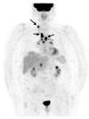
Caucasian woman (78 years old) suffering from bilateral unexplained panuveitis. Whole body 18F-FDG PET (maximum intensity projection anterior view) demonstrated significant bilateral hilar, mediastinal and right supraclavicular lymph nodes uptakes (black arrows) while there was no node enlargement on chest CT. A subsequent supraclavicular node dissection revealed noncaseating epithelioid granulomas consistent with sarcoidosis. Special staining and culture for mycobacteria were negative.
Figure 10
Figure 10
Lung biopsy: non-necrotizing epithelioid granulomas (arrows) with giant cells surrounding lymphocytes and fibrosis.

References

    1. Besnier E.H. Lupus Pernio de La Face; Synovites Fongueuses (Scrofulo-Tuberculeuses) Symétriques Des Extrémités Supérieures. Ann. Derm. Syph. 1889;10:333–336.
    1. Sakthivel P., Bruder D. Mechanism of Granuloma Formation in Sarcoidosis. Curr. Opin. Hematol. 2017;24:59–65. doi: 10.1097/MOH.0000000000000301. - DOI - PubMed
    1. Hunninghake G., Costabel U. Statement on Sarcoidosis. Am. J. Respir. Crit. Care Med. 1999;160:20. doi: 10.1034/j.1399-3003.1999.14d02.x. - DOI
    1. Arkema E.V., Cozier Y.C. Epidemiology of Sarcoidosis: Current Findings and Future Directions. Ther. Adv. Chronic Dis. 2018;9:227–240. doi: 10.1177/2040622318790197. - DOI - PMC - PubMed
    1. Rybicki B.A., Major M., Popovich J., Maliank M.J., Lannuzzi M.C. Racial Differences in Sarcoidosis Incidence: A 5-Year Study in a Health Maintenance Organization. Am. J. Epidemiol. 1997;145:234–241. doi: 10.1093/oxfordjournals.aje.a009096. - DOI - PubMed

LinkOut - more resources