

Beat Detection Recruits the Visual Cortex in Early Blind Subjects
- PMID: 33807372
- PMCID: PMC8066101
- DOI: 10.3390/life11040296
Beat Detection Recruits the Visual Cortex in Early Blind Subjects
Abstract
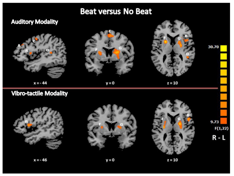
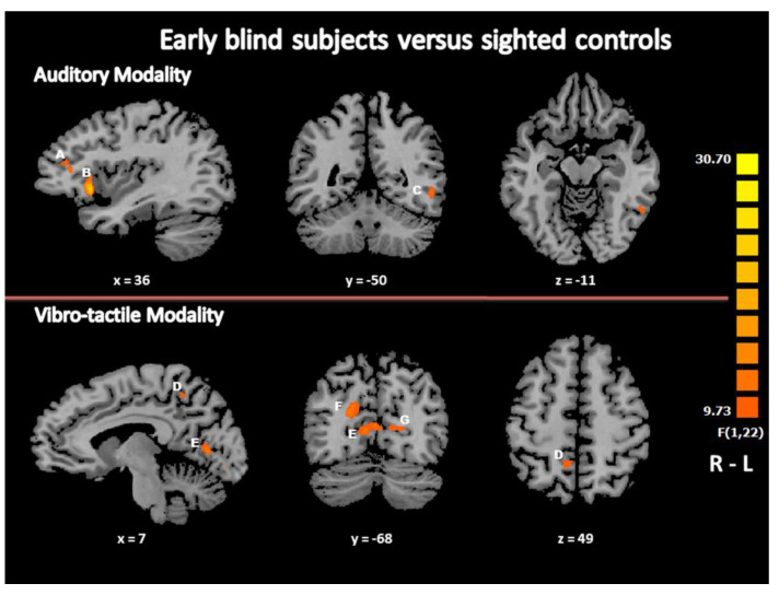
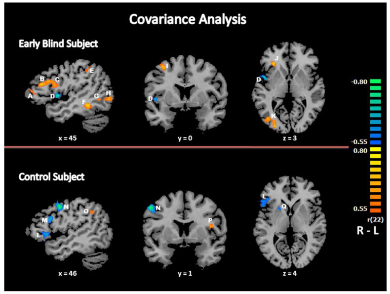
Using functional magnetic resonance imaging, here we monitored the brain activity in 12 early blind subjects and 12 blindfolded control subjects, matched for age, gender and musical experience, during a beat detection task. Subjects were required to discriminate regular ("beat") from irregular ("no beat") rhythmic sequences composed of sounds or vibrotactile stimulations. In both sensory modalities, the brain activity differences between the two groups involved heteromodal brain regions including parietal and frontal cortical areas and occipital brain areas, that were recruited in the early blind group only. Accordingly, early blindness induced brain plasticity changes in the cerebral pathways involved in rhythm perception, with a participation of the visually deprived occipital brain areas whatever the sensory modality for input. We conclude that the visually deprived cortex switches its input modality from vision to audition and vibrotactile sense to perform this temporal processing task, supporting the concept of a metamodal, multisensory organization of this cortex.
Keywords: beat perception; early blindness; multisensory integration; rhythm perception; touch; vision.
Conflict of interest statement
The authors declare no conflict of interest.
Figures

Similar articles
-
Hearing, feeling or seeing a beat recruits a supramodal network in the auditory dorsal stream.Eur J Neurosci. 2017 Jun;45(11):1439-1450. doi: 10.1111/ejn.13349. Epub 2016 Aug 12. Eur J Neurosci. 2017. PMID: 27471102
-
Improved beat asynchrony detection in early blind individuals.Perception. 2014;43(10):1083-96. doi: 10.1068/p7789. Perception. 2014. PMID: 25509685
-
Whole brain functional connectivity in the early blind.Brain. 2007 Aug;130(Pt 8):2085-96. doi: 10.1093/brain/awm121. Epub 2007 May 28. Brain. 2007. PMID: 17533167
-
The Cross-Modal Effects of Sensory Deprivation on Spatial and Temporal Processes in Vision and Audition: A Systematic Review on Behavioral and Neuroimaging Research since 2000.Neural Plast. 2019 Dec 2;2019:9603469. doi: 10.1155/2019/9603469. eCollection 2019. Neural Plast. 2019. PMID: 31885540 Free PMC article.
-
Compensatory plasticity and cross-modal reorganization following early visual deprivation.Neurosci Biobehav Rev. 2014 Apr;41:36-52. doi: 10.1016/j.neubiorev.2013.08.001. Epub 2013 Aug 15. Neurosci Biobehav Rev. 2014. PMID: 23954750 Review.
Cited by
-
Grey Matter Hypertrophy and Atrophy in Early-Blind Adolescents: A Surface-Based Morphometric Study.Dis Markers. 2022 May 3;2022:8550714. doi: 10.1155/2022/8550714. eCollection 2022. Dis Markers. 2022. PMID: 35557871 Free PMC article.
References
Grants and funding
LinkOut - more resources
Full Text Sources
Other Literature Sources
