. 2021 Mar 31;22(7):3646.
doi: 10.3390/ijms22073646.

Long-Term Iron Deficiency and Dietary Iron Excess Exacerbate Acute Dextran Sodium Sulphate-Induced Colitis and Are Associated with Significant Dysbiosis

Affiliations

Long-Term Iron Deficiency and Dietary Iron Excess Exacerbate Acute Dextran Sodium Sulphate-Induced Colitis and Are Associated with Significant Dysbiosis

Awad Mahalhal et al. Int J Mol Sci. .

Abstract

Background: Oral iron supplementation causes gastrointestinal side effects. Short-term alterations in dietary iron exacerbate inflammation and alter the gut microbiota, in murine models of colitis. Patients typically take supplements for months. We investigated the impact of long-term changes in dietary iron on colitis and the microbiome in mice.

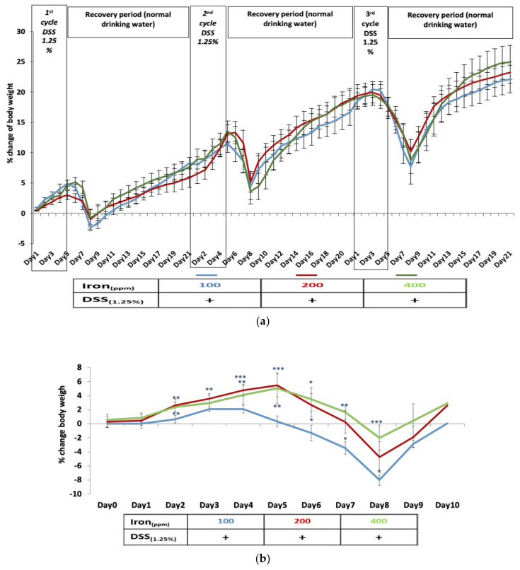
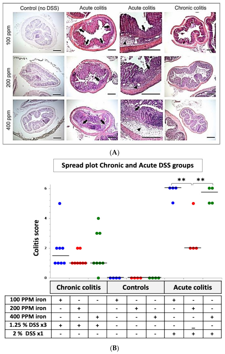
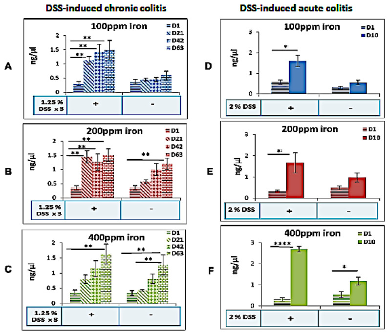
Methods: We fed mice chow containing differing levels of iron, reflecting deficient (100 ppm), normal (200 ppm), and supplemented (400 ppm) intake for up to 9 weeks, both in absence and presence of dextran sodium sulphate (DSS)-induced chronic colitis. We also induced acute colitis in mice taking these diets for 8 weeks. Impact was assessed (i) clinically and histologically, and (ii) by sequencing the V4 region of 16S rRNA.

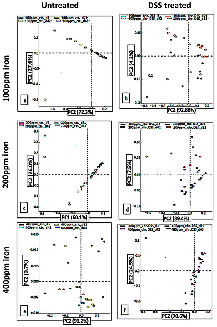
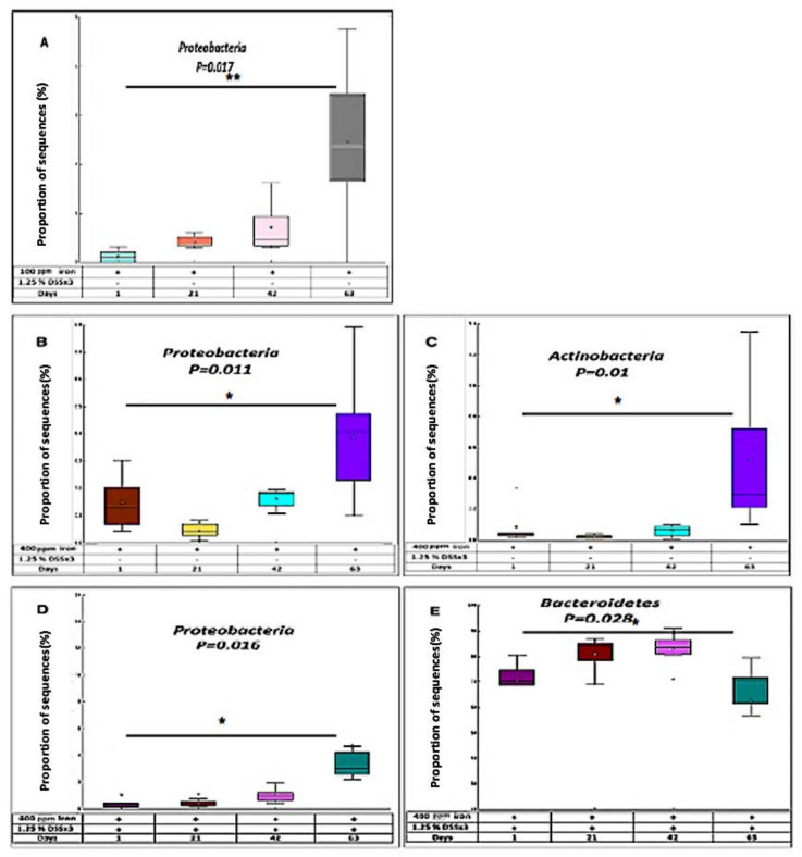
Results: In mice with long-term changes, the iron-deficient diet was associated with greater weight loss and histological inflammation in the acute colitis model. Chronic colitis was not influenced by altering dietary iron however there was a change in the microbiome in DSS-treated mice consuming 100 ppm and 400 ppm iron diets, and control mice consuming the 400 ppm iron diet. Proteobacteria levels increased significantly, and Bacteroidetes levels decreased, in the 400 ppm iron DSS group at day-63 compared to baseline.

Conclusions: Long-term dietary iron alterations affect gut microbiota signatures but do not exacerbate chronic colitis, however acute colitis is exacerbated by such dietary changes. More work is needed to understand the impact of iron supplementation on IBD. The change in the microbiome, in patients with colitis, may arise from the increased luminal iron and not simply from colitis.

Keywords: diet; fecal microbiota; inflammatory bowel disease; intestinal inflammation; iron.


Conflict of interest statement

The authors have declared that no competing interest exists. The funder had no role in study design, data collection and analysis, decision to publish, nor in preparation of the manuscript.

Figures

Figure 1
Figure 1
Dextran sulfate sodium (DSS) colitis – induced body weight changes in mice consuming diets of differing dietary iron levels. (a) Percentage weight loss observed in mice with chronic colitis induced by three cycles of 1.25% w/v DSS during the 63-day period was not influenced by consumption of an iron-deficient diet (100 ppm iron [blue]), standard chow diet (200 ppm iron [red]) nor an iron supplemented diet (400 ppm iron [green]); Data presented as mean ± standard error of the mean (SEM). No statistical differences were seen between groups (n = 8 mice per group). (b) Percentage body weight loss in mice resulting from DSS-induced acute colitis on 63 days on deficient and supplemented iron diets compared with standard chow (n = 4 female mice per group). Statistical differences * p < 0.05, ** p < 0.01, *** p < 0.001; Kruskal–Wallis test followed by multiple comparison tests.
Figure 2
Figure 2
Histological analysis of colon from DSS-treated mice in consuming diets of differing dietary iron levels. (A) Representative hematoxylin and eosin-stained segments of distal colon from control C57BL/6J mice ingesting 100, 200 or 400 ppm iron diets alone (n = 4 mice in total), C57BL/6 with acute (n = 4) and chronic DSS-induced colitis (n = 8) administered with 100, 200 or 400 ppm iron diets as indicated. Arrowheads highlight submucosal oedema; arrows highlight almost complete loss of colonic epithelium and leukocyte infiltration. Scale bar: 200 µm. (B): Inflammation (colitis) scores for all groups’ DSS-treated (24 (63-days) and 12 (10-days) mice per group) and untreated (controls) 12 (63-days) mice on different iron diets. Horizontal lines at the median. an iron deficient diet (100 ppm iron [blue]), standard chow diet (200 ppm iron [red]) nor an iron supplemented diet (400 ppm iron [green]) Differences tested by Kruskal–Wallis test followed by multiple comparison tests ** p < 0.01.
Figure 3
Figure 3
Fecal iron changes in mice fed differing iron diets in the presence or absence of colitis. Fecal iron concentrations in the presence or absence of DSS-induced chronic DSS (day-1 vs. day-21, day-42, and day-63; n = 8 mice per group) or acute colitis DSS (day-1 vs. day-10; n = 4 mice per group), for mice consuming a (A,D) deficient (100 ppm iron), (B,E) standard (200 ppm iron) or (C,F) supplemented (400 ppm iron) chow diet respectively. Data are presented as a mean ± standard error of the mean (SEM). Differences were tested by Kruskal–Wallis test followed by multiple comparison Dunn’s test; * p < 0.05, ** p < 0.01, **** p < 0.0001.
Figure 4
Figure 4
PCA to show unweighted UniFrac diseases after DSS treatment. In chronic DSS, PCA plots of the unweighted UniFrac distances of pre-and post-DSS-intervention stool samples from chronic (3 cycles) DSS – treated mice (b,d,f) and (a,c,e) untreated mice at phylum-level, phylogenetic classification of 16S rRNA gene sequences. Symbols represent data from individual mice, color-coded by the indicated metadata. Statistical differences were assessed by Kruskal–Wallis H-test followed by Storey’s FDR multiple test correction.
Figure 5
Figure 5
Distribution of bacteria in response to chronic DSS. (A) In chronic DSS, box plot showing the distribution in the proportion of Proteobacteria assigned to samples at day-1, 21, 42, and 63 from 100 ppm iron DSS-treated mice. In chronic DSS, box plot showing the distribution in the proportion of two phyla (Proteobacteria (B) and Actinobacteria (C)) assigned to samples from 400 ppm iron untreated mice. In chronic DSS, box plot showing the distribution in the proportion of two phyla (Proteobacteria (D) and Bacteroidetes (E)) assigned to samples from 400 ppm iron DSS-treated mice.

References

    1. Kaser A., Zeissig S., Blumberg R.S. Inflammatory bowel disease. Annu. Rev. Immunol. 2010;28:573–621. doi: 10.1146/annurev-immunol-030409-101225. - DOI - PMC - PubMed
    1. Constante M., Fragoso G., Lupien-Meilleur J., Calvé A., Santos M.M. Iron Supplements Modulate Colon Microbiota Composition and Potentiate the Protective Effects of Probiotics in Dextran Sodium Sulfate-induced Colitis. Inflamm. Bowel Dis. 2017;23:753–766. doi: 10.1097/MIB.0000000000001089. - DOI - PubMed
    1. Mowat C., Cole A., Windsor A., Ahmad T., Arnott I., Driscoll R., Mitton S., Orchard T., Rutter M., Younge L., et al. Guidelines for the management of inflammatory bowel disease in adults. Gut. 2011;60:571–607. doi: 10.1136/gut.2010.224154. - DOI - PubMed
    1. Manfred Wick W.P., Lehmann P. Clinical Aspects and Laboratory—Iron Metabolism. 6th ed. Springer; Vienna, Austria: 2011.
    1. Kulnigg S., Teischinger L., Dejaco C., Waldhör T., Gasche C. Rapid Recurrence of IBD-Associated Anemia and Iron Deficiency After Intravenous Iron Sucrose and Erythropoietin Treatment. Am. J. Gastroenterol. 2009;104:1460–1467. doi: 10.1038/ajg.2009.114. - DOI - PubMed

MeSH terms

LinkOut - more resources