. 2021 Mar 31;9(4):735.
doi: 10.3390/microorganisms9040735.

Peroral Clove Essential Oil Treatment Ameliorates Acute Campylobacteriosis-Results from a Preclinical Murine Intervention Study

Affiliations

Peroral Clove Essential Oil Treatment Ameliorates Acute Campylobacteriosis-Results from a Preclinical Murine Intervention Study

Stefan Bereswill et al. Microorganisms. .

Abstract

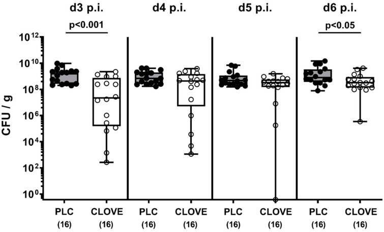
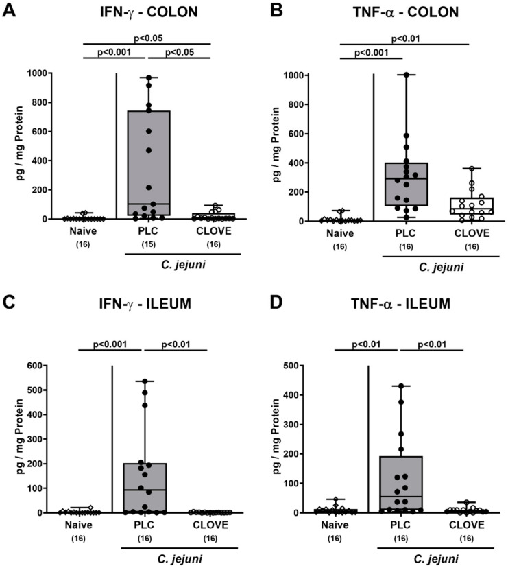
Campylobacter (C.) jejuni infections pose progressively emerging threats to human health worldwide. Given the rise in antibiotic resistance, antibiotics-independent options are required to fight campylobacteriosis. Since the health-beneficial effects of clove have been known for long, we here analyzed the antimicrobial and immune-modulatory effects of clove essential oil (EO) during acute experimental campylobacteriosis. Therefore, microbiota-depleted interleukin-10 deficient (IL-10-/-) mice were perorally infected with C. jejuni and treated with clove EO via drinking water starting on day 2 post-infection. On day 6 post-infection, lower small- and large-intestinal pathogen loads could be assessed in clove EO as compared to placebo treated mice. Although placebo mice suffered from severe campylobacteriosis as indicated by wasting and bloody diarrhea, clove EO treatment resulted in a better clinical outcome and in less severe colonic histopathological and apoptotic cell responses in C. jejuni infected mice. Furthermore, lower colonic numbers of macrophages, monocytes, and T lymphocytes were detected in mice from the verum versus the placebo cohort that were accompanied by lower intestinal, extra-intestinal, and even systemic proinflammatory cytokine concentrations. In conclusion, our preclinical intervention study provides first evidence that the natural compound clove EO constitutes a promising antibiotics-independent treatment option of acute campylobacteriosis in humans.

Keywords: Campylobacter jejuni; acute campylobacteriosis model; clove essential oil; enteropathogenic infection; eugenol; host-pathogen interaction; immune-modulatory effects; microbiota-depleted IL-10−/− mice; natural antibiotics-independent compounds; preclinical intervention study.


Conflict of interest statement

The funders had no role in the design of the study; in the collection, analyses, or interpretation of data; in the writing of the manuscript, or in the decision to publish the results.

Figures

Figure 1
Figure 1
Kinetic survey of fecal pathogen burdens following clove essential oil (EO) treatment of Campylobacter (C.) jejuni infected interleukin-10 deficient (IL-10−/−) mice. Microbiota-depleted IL-10−/− mice were perorally infected with C. jejuni strain 81-176 on day (d) 0 and d1. From d2 until d6 post-infection (p.i.), mice were perorally challenged with clove EO or received placebo (PLC) via drinking water. The pathogen numbers were determined in fecal samples over time by culture and expressed as colony forming units (CFU) per g). Box plots indicate the 75th and the 25th percentiles of the median (black bar within box). The total range, significance levels (p values) determined by the Mann–Whitney U test and the total numbers of analyzed mice (in parentheses) are given. Data were pooled from four independent experiments.
Figure 2
Figure 2
Gastrointestinal pathogen burdens following clove EO treatment of C. jejuni infected IL-10−/− mice. Microbiota-depleted IL-10−/− mice were perorally infected with C. jejuni strain 81-176 on day (d) 0 and d1. From d2 until d6 post-infection, mice were perorally challenged with clove EO or received placebo (PLC) via drinking water. On d6 post-infection, the pathogen numbers were determined in luminal samples taken from distinct gastrointestinal parts by culture and expressed as colony forming units per g, CFU/g). Box plots indicate the 75th and the 25th percentiles of the median (black bar within box). The total range, significance levels (p values) determined by the Mann–Whitney U test and the number of pathogen-positive out of the total number of analyzed samples (in parentheses) are given. Data were pooled from four independent experiments.
Figure 3
Figure 3
Kinetic survey of C. jejuni induced clinical conditions following clove EO treatment of infected IL-10−/− mice. Microbiota-depleted IL-10−/− mice were perorally infected with C. jejuni strain 81-176 on day (d) 0 and d1. From d2 until d6 post-infection (p.i.), mice were perorally challenged with clove EO or received placebo (PLC) via drinking water. The clinical conditions of mice were quantitatively determined over time, i.e., on (A) d3, (B) d4, (C) d5 and (D) d6 p.i. by using a clinical scoring system by using a clinical scoring system. Box plots indicate the 75th and the 25th percentiles of the median (black bar within box). Naive mice were included as negative control animals. The total range, significance levels (p values) determined by the Kruskal-Wallis test and Dunn’s post-correction and the number of mice with clinical signs out of the total number of analyzed animals (in parentheses) are given. Data were pooled from four independent experiments.
Figure 4
Figure 4
C. jejuni induced microscopic inflammatory sequelae following clove EO treatment of infected IL-10−/− mice. Microbiota-depleted IL-10−/− mice were perorally infected with C. jejuni strain 81-176 on day (d) 0 and d1. From d2 until d6 post-infection (p.i.), mice were perorally challenged with clove EO or received placebo (PLC) via drinking water. On day 6 p.i., (A) colonic histopathological changes were quantified in hematoxylin and eosin-stained colonic paraffin sections by using histopathological scores. Furthermore, (B) the average numbers of apoptotic colonic epithelial cells were assessed microscopically from six high power fields (HPF, 400 × magnification) per animal in paraffin sections positive for cleaved caspase3 (Casp3+). Box plots indicate the 75th and the 25th percentiles of the median (black bar within box). Naive mice were included as negative control animals. The total range, significance levels (p values) determined by the ANOVA test with Tukey post-correction or by the Kruskal-Wallis test and Dunn’s post-correction and the total numbers of analyzed mice (in parentheses) are given. Definite outliers were removed after being identified by the Grubb’s test (α = 0.001). Data were pooled from four independent experiments.
Figure 5
Figure 5
C. jejuni induced immune cell responses following clove EO treatment of infected IL-10−/− mice. Microbiota-depleted IL-10−/− mice were perorally infected with C. jejuni strain 81-176 on day (d) 0 and d1. From d2 until d6 post-infection (p.i.), mice were perorally challenged with clove EO or received placebo (PLC) via drinking water. On day 6 p.i., the average numbers of (A) macrophages and monocytes (F4/80+), (B) T lymphocytes (CD3+), (C) regulatory T cells (FOXP3+) and (D) B lymphocytes (B220+) per animal were determined in immunohistochemically stained colonic paraffin sections from six high power fields (HPF, 400× magnification). Box plots indicate the 75th and the 25th percentiles of the median (black bar within box). Naive mice were included as negative control animals. The total range, significance levels (p values) determined by the Kruskal-Wallis test and Dunn’s post-correction and the total numbers of analyzed mice (in parentheses) are given. Data were pooled from four independent experiments.
Figure 6
Figure 6
C. jejuni induced intestinal proinflammatory cytokine secretion following clove EO treatment of infected IL-10−/− mice. Microbiota-depleted IL-10−/− mice were perorally infected with C. jejuni strain 81-176 on day (d) 0 and d1. From d2 until d6 post-infection (p.i.), mice were perorally challenged with clove EO or received placebo (PLC) via drinking water. On day 6 p.i., (A,C) IFN-γ and (B,D) TNF-α concentrations were measured were measured in ex vivo biopsies derived from the (A,B) colon and (C,D) ileum. Box plots indicate the 75th and the 25th percentiles of the median (black bar within box). Naive mice were included as negative control animals. The total range, significance levels (p values) determined by the Kruskal-Wallis test and Dunn’s post-correction and the total numbers of analyzed mice (in parentheses) are given. Data were pooled from four independent experiments.
Figure 7
Figure 7
C. jejuni induced extra-intestinal proinflammatory cytokine secretion following clove EO treatment of infected IL-10−/− mice. Microbiota-depleted IL-10−/− mice were perorally infected with C. jejuni strain 81-176 on day (d) 0 and d1. From d2 until d6 post-infection (p.i.), mice were perorally challenged with clove EO or received placebo (PLC) via drinking water. On day 6 p.i., (A,C) IFN-γ and (B,D) TNF-α concentrations were measured in ex vivo biopsies derived from the (A,B) liver and (C,D) kidneys. Box plots indicate the 75th and the 25th percentiles of the median (black bar within box). Naive mice were included as negative control animals. The total range, significance levels (p values) determined by the Kruskal-Wallis test and Dunn’s post-correction and the total numbers of analyzed mice (in parentheses) are given. Data were pooled from four independent experiments.
Figure 8
Figure 8
C. jejuni induced splenic proinflammatory mediator secretion following clove EO treatment of infected IL-10−/− mice. Microbiota-depleted IL-10−/− mice were perorally infected with C. jejuni strain 81-176 on day (d) 0 and d1. From d2 until d6 post-infection (p.i.), mice were perorally challenged with clove EO or received placebo (PLC) via drinking water. On day 6 p.i., (A) IFN-γ, (B) TNF-α, (C) MCP-1 and (D) IL-6 concentrations were measured in ex vivo biopsies derived from the spleen. Box plots indicate the 75th and the 25th percentiles of the median (black bar within box). Naive mice were included as negative control animals. The total range, significance levels (p values) determined by the Kruskal-Wallis test and Dunn’s post-correction and the total numbers of analyzed mice (in parentheses) are given. Data were pooled from four independent experiments.
Figure 9
Figure 9
C. jejuni induced systemic proinflammatory mediator secretion following clove EO treatment of infected IL-10−/− mice. Microbiota-depleted IL-10−/− mice were perorally infected with C. jejuni strain 81-176 on day (d) 0 and d1. From d2 until d6 post-infection (p.i.), mice were perorally challenged with clove EO or received placebo (PLC) via drinking water. On day 6 p.i., (A) IFN-γ, (B) TNF-α, (C) MCP-1 and (D) IL-6 concentrations were measured in serum samples. Box plots indicate the 75th and the 25th percentiles of the median (black bar within box). Naive mice were included as negative control animals. The total range, significance levels (p values) determined by the Kruskal-Wallis test and Dunn’s post-correction and the total numbers of analyzed mice (in parentheses) are given. Definite outliers were removed after being identified by the Grubb’s test (α = 0.001). Data were pooled from four independent experiments.

Similar articles

Cited by

References

    1. Kaakoush N.O., Castano-Rodriguez N., Mitchell H.M., Man S.M. Global Epidemiology of Campylobacter Infection. Clin. Microbiol. Rev. 2015;28:687–720. doi: 10.1128/CMR.00006-15. - DOI - PMC - PubMed
    1. WHO World Health Organisation. Campylobacter. [(accessed on 4 June 2020)]; Available online: https://www.who.int/news-room/fact-sheets/detail/campylobacter.
    1. EFSA European Food Safety Authority. European Centre for Disease, Prevention Control, The European Union One Health 2018 Zoonoses Report. EFSA J. 2019;17:e05926. doi: 10.2903/j.efsa.2019.5926. - DOI - PMC - PubMed
    1. Heimesaat M.M., Backert S., Alter T., Bereswill S. Human Campylobacteriosis-A Serious Infectious Threat in a One Health Perspective. Curr. Top. Microbiol. Immunol. 2021;431:1–23. - PubMed
    1. Young K.T., Davis L.M., Dirita V.J. Campylobacter jejuni: Molecular biology and pathogenesis. Nat. Rev. Microbiol. 2007;5:665–679. doi: 10.1038/nrmicro1718. - DOI - PubMed

LinkOut - more resources