Review
. 2021 Mar 5;10(3):562.
doi: 10.3390/cells10030562.

Towards a Framework for Better Understanding of Quiescent Cancer Cells

Affiliations
Review

Towards a Framework for Better Understanding of Quiescent Cancer Cells

Wan Najbah Nik Nabil et al. Cells. .

Abstract

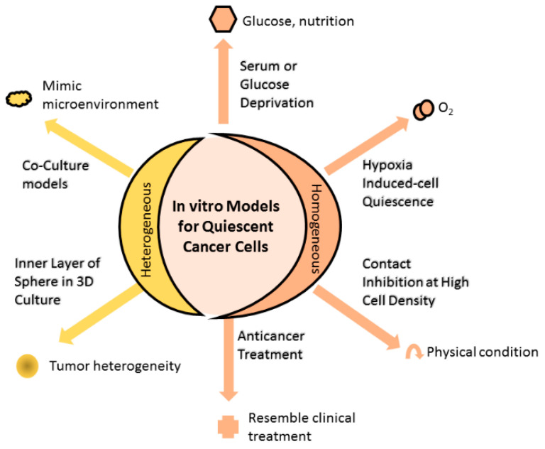
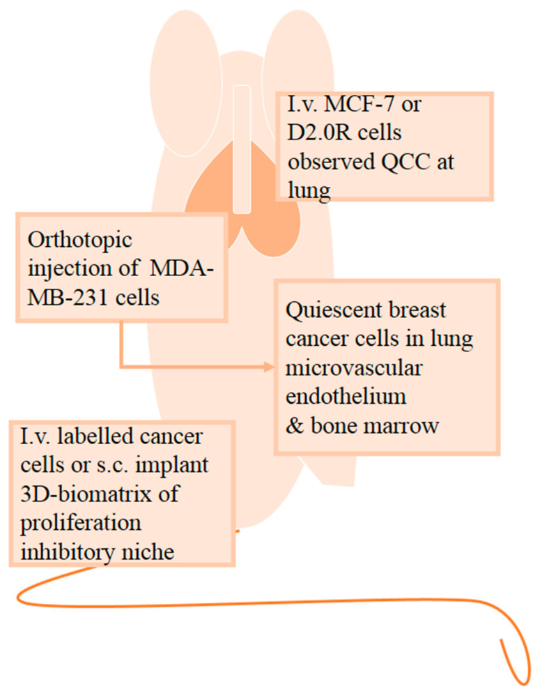
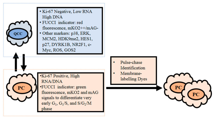
Quiescent cancer cells (QCCs) are cancer cells that are reversibly suspended in G0 phase with the ability to re-enter the cell cycle and initiate tumor growth, and, ultimately, cancer recurrence and metastasis. QCCs are also therapeutically challenging due to their resistance to most conventional cancer treatments that selectively act on proliferating cells. Considering the significant impact of QCCs on cancer progression and treatment, better understanding of appropriate experimental models, and the evaluation of QCCs are key questions in the field that have direct influence on potential pharmacological interventions. Here, this review focuses on existing and emerging preclinical models and detection methods for QCCs and discusses their respective features and scope for application. By providing a framework for selecting appropriate experimental models and investigative methods, the identification of the key players that regulate the survival and activation of QCCs and the development of more effective QCC-targeting therapeutic agents may mitigate the consequences of QCCs.

Keywords: cancer; detection; dormancy; model; quiescence.


Conflict of interest statement

The authors declare no conflict of interest.

Figures

Figure 1
Figure 1
In vitro models for quiescent cancer cells (QCCs). Changing the physical condition of cell culture by allowing cultured cells to grow at high cell density until reaching contact inhibition or depriving essential substances (such as serum, glucose, or oxygen) of the culture, or administering anticancer treatment into cell culture that homogenously induces cells into quiescence. Alternatively, cell culture can heterogeneously comprise quiescent and proliferating cells using the 3D culture method. The inner and outer layers of 3D spheres are mainly composed of quiescent and proliferating cells, respectively, resembling the tumor heterogeneity observed in the clinical setting. Other means to achieve heterogeneous cell composition are coculturing different cell populations that mimic the microenvironment in which actual tumor cells grow, while anticancer treatment induces a portion of cells to quiescence, as found in the clinical setting.
Figure 2
Figure 2
In vivo quiescent models are applied to examine the interactions between cancer cells and the systemic host response. To study the dissemination of quiescent cancer cells (QCCs) to metastatic sites, namely, the lung, mice were intravenously (i.v.) injected with breast cancer MCF-7 cells or mouse mammary carcinoma D2.0R cells. The effect of the environmental niche on QCCs can be examined by either intravenous injection of labeled cancer cells or subcutaneously (s.c.) implanting 3D biomatrix that serves as a proliferation inhibitory niche. Following orthotopic injection of MDA-MB-231 breast cancer cells into mice, QCCs were detected in the microvascular endothelium of the lung and bone marrow.
Figure 3
Figure 3
Current detection methodology for quiescent cancer cells (QCCs) that reside at G0 phase. QCCs are generally detected based on their Ki-67 negative and low cellular RNA content, while proliferating cells are Ki-67 positive and contain high RNA and DNA. Other detection methods are also applied to assist in differentiating between quiescent and proliferating cells. With a fluorescence ubiquitin cell cycle indicator (FUCCI), G0/G1 cells are labeled with red fluorescence or identified based on their mKO2++/mAG- signal. Proliferating cells are identifiable by FUCCI as green fluorescence and further distinguished via mKO2 and mAG signals: G1 (mKO2+/mAG-), very early G1 (mKO2-/mAG-), G1/S (mKO2+/mAG+), and S/G2/M (mKO2-/mAG+) phases. Other potential molecular markers for QCCs are p38high/ERKlow, MCM2low/H3K9me2low/HES1high; high levels of p27, dual specificity tyrosine phosphorylation-regulated kinase 1B (DYRK1B), nuclear receptor subfamily 2 group F member 1 (NR2F1), and G0/G1 switch gene 2 (GOS2); and low levels of c-Myc and reactive oxygen species (ROS). Pulse-chase identification and membrane-labeling dye methods involve dilution of respective fluorescent and membrane-labeling dyes as cells proliferate. Hence, label-retaining cells are slow cycling cells, while label-diluted cells are proliferating cells.

References

    1. Phan T.G., Croucher P.I. The dormant cancer cell life cycle. Nat. Rev. Cancer. 2020;20:398–411. doi: 10.1038/s41568-020-0263-0. - DOI - PubMed
    1. National Cancer Comprehensive Network (NCCN) Breast Cancer, Version 4.2020, NCCN Clinical Practice Guidelines in Oncology. [(accessed on 16 June 2020)]; Available online: https://www.nccn.org/professionals/physician_gls/pdf/breast.pdf.
    1. Yao G. Modelling mammalian cellular quiescence. Interface Focus. 2014;4:34458–34474. doi: 10.1098/rsfs.2013.0074. - DOI - PMC - PubMed
    1. Endo H., Inoue M. Dormancy in cancer. Cancer Sci. 2019;110:474–480. doi: 10.1111/cas.13917. - DOI - PMC - PubMed
    1. Pack L.R., Daigh L.H., Meyer T. Putting the brakes on the cell cycle: Mechanisms of cellular growth arrest. Curr. Opin. Cell Biol. 2019;60:106–113. doi: 10.1016/j.ceb.2019.05.005. - DOI - PMC - PubMed

Publication types

LinkOut - more resources