. 2021 Mar 5;22(5):2614.
doi: 10.3390/ijms22052614.

CNBP Binds and Unfolds In Vitro G-Quadruplexes Formed in the SARS-CoV-2 Positive and Negative Genome Strands

Affiliations

CNBP Binds and Unfolds In Vitro G-Quadruplexes Formed in the SARS-CoV-2 Positive and Negative Genome Strands

Georgina Bezzi et al. Int J Mol Sci. .

Abstract

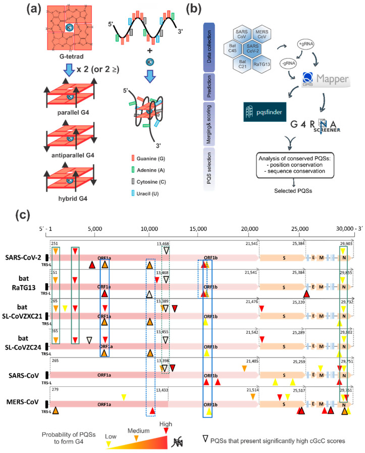
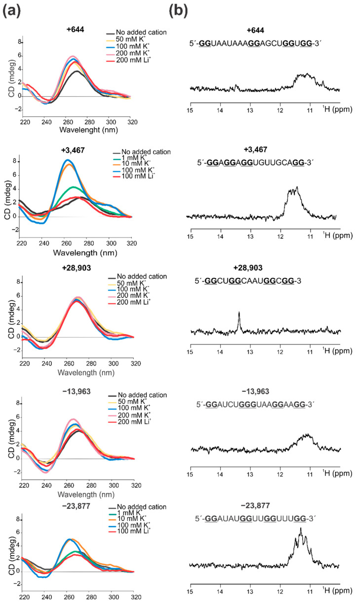
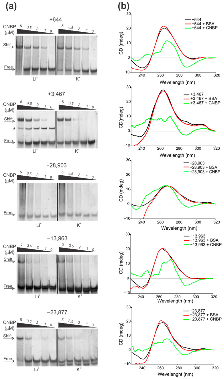
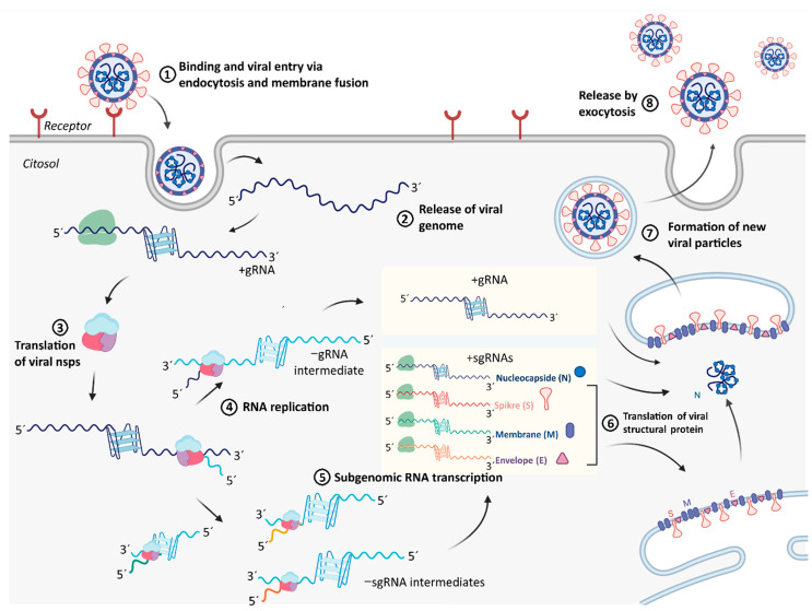
The Coronavirus Disease 2019 (COVID-19) pandemic has become a global health emergency with no effective medical treatment and with incipient vaccines. It is caused by a new positive-sense RNA virus called severe acute respiratory syndrome-related coronavirus 2 (SARS-CoV-2). G-quadruplexes (G4s) are nucleic acid secondary structures involved in the control of a variety of biological processes including viral replication. Using several G4 prediction tools, we identified highly putative G4 sequences (PQSs) within the positive-sense (+gRNA) and negative-sense (-gRNA) RNA strands of SARS-CoV-2 conserved in related betacoronaviruses. By using multiple biophysical techniques, we confirmed the formation of two G4s in the +gRNA and provide the first evidence of G4 formation by two PQSs in the -gRNA of SARS-CoV-2. Finally, biophysical and molecular approaches were used to demonstrate for the first time that CNBP, the main human cellular protein bound to SARS-CoV-2 RNA genome, binds and promotes the unfolding of G4s formed by both strands of SARS-CoV-2 RNA genome. Our results suggest that G4s found in SARS-CoV-2 RNA genome and its negative-sense replicative intermediates, as well as the cellular proteins that interact with them, are relevant factors for viral genes expression and replication cycle, and may constitute interesting targets for antiviral drugs development.

Keywords: CNBP; COVID-19; G-quadruplex; SARS-CoV-2; coronavirus.


Conflict of interest statement

The authors declare no conflict of interest. The funders had no role in the design of the study; in the collection, analyses, or interpretation of data; in the writing of the manuscript, or in the decision to publish the results.

Figures

Figure 1
Figure 1
Identification and selection of PQSs in the genomes of SARS-CoV-2 and other members of the Coronaviridae family. (a) Cartoon representing the formation of G4s. Left: G-tetrads are formed by the planar arrange of four G nucleobases stabilized by lateral Hoogsteen-type hydrogen bonds. The stacking of two or more tetrads and the coordination of K+ form the G4 structure. Depending on the relative orientation of the G-tracts, G4s may be parallel, antiparallel, or hybrid. Right: G4 formation by the folding on itself of a G-rich RNA strand with at least four contiguous G-tracts interspersed with short nucleotide loops. The formation of a parallel G4 is represented. (b) Schematic summary of the bioinformatic workflow conducted for PQSs identification and selection in the positive- and negative-sense RNA genomes of SARS-CoV-2, RaTG13, bat-SL-CoVZC45, bat-SL-CoVZXC21, SARS-CoV, and MERS-CoV. (c) Schematic representation of the location of the selected PQSs in the analyzed genomes. The organization of the coding regions for the main viral proteins are represented for each virus: ORF1a (open-reading frame 1a), ORF1b (open-reading frame 1b), S (spike), E (envelope), M (membrane) and N (nucleocapsid), black rectangle represents transcription-regulatory sequences (TRS) in the 5′ untranslated region (UTR). PQSs found in the positive-sense strand are represented above the genome while PQSs found on the negative-sense strand represented below the genome. PQSs are represented with the following color code: PQSs with high probability to form G4 (red), PQSs with medium probability to form G4 (orange) and PQSs with low probability to form G4 (yellow). PQSs that present significantly high cGcC scores (>150) are highlighted using thick edges. PQSs conserved in position and sequence are indicated with solid green line boxes (for positive-sense strand) and solid blue line boxes (for negative-sense strand). Dashed lines indicate PQSs conserved in position that were not selected due to lack of sequence conservation, to conservation in less than four genomes or to not overpassing the selection criterion.
Figure 2
Figure 2
Evidence by CD and NMR spectroscopy of the in vitro G4 structures formed by the selected PQSs. (a) CD spectra were obtained for each RNA sequence (named by the PQS position) folded in the absence and in the presence of increasing K+ concentrations, or in the presence of Li+ at the highest concentration used for K+. Concentrations are indicated for each plot. (b) 1D 1H NMR spectra obtained for each RNA sequence (named by the PQS position) folded in the presence of K+ at the highest concentration used for CD. RNA sequence for each PQS are represented above NMR spectra, and guanine nucleotides predicted to participate in the G4 formation are indicated in bold and underlined.
Figure 3
Figure 3
CNBP binds and unfolds the G4s formed by PQSs in the +gRNA and −gRNA of SARS-CoV-2. (a) Representative EMSAs performed using PQSs (named by the PQS position) as probes folded in the presence of Li+ (left) or K+ (right) and then incubated in the absence or presence of increasing concentrations of CNBP. Free and shifted probes are indicated by arrows at the left of the gels. The +3467 probe folded in the presence of Li+ presents a minority band (marked with *) of lower mobility probably due to a self-assembled dimeric or multimeric complex. (b) CD spectra obtained for 8 µM oligonucleotides (named by the PQS position) folded as G4 in the presence of K+ and incubated in the absence of protein or in the presence of CNBP (1:1 molar ratio) or BSA. For EMSAs and CD, K+ and Li+ concentrations used for G4 folding were 100 mM (+3467 and −23,877) or 200 mM (+644, +28,903 and −13,963).
Figure 4
Figure 4
Possible role of G4s in the SARS-CoV-2 and coronaviruses replication cycle. Different steps of viral infection and replicative cycle are detailed: (1) binding to host cell receptor, (2) entry to host cell, (3) translation of viral nsps from ORF1ab, (4) viral RNA replication, (5) viral RNA transcription, (6) viral structural proteins translation, (7) encapsidation of viral +gRNA and formation of mature virus particles, (8) viral release. Viral G4s and the G4-interacting proteins, such as CNBP, may participate in the regulation of the efficiency of steps 3–7, which could be targets of drug candidates for antiviral therapies. Created with BioRender.com.

References

    1. Zhou P., Yang X.L., Wang X.G., Hu B., Zhang L., Zhang W., Si H.R., Zhu Y., Li B., Huang C.L., et al. A pneumonia outbreak associated with a new coronavirus of probable bat origin. Nature. 2020;579:270–273. doi: 10.1038/s41586-020-2012-7. - DOI - PMC - PubMed
    1. Gorbalenya A.E., Baker S.C., Baric R.S., de Groot R.J., Drosten C., Gulyaeva A.A., Haagmans B.L., Lauber C., Leontovich A.M., Neuman B.W., et al. The species Severe acute respiratory syndrome-related coronavirus: Classifying 2019-nCoV and naming it SARS-CoV-2. Nat. Microbiol. 2020;5:536–544. doi: 10.1038/s41564-020-0695-z. - DOI - PMC - PubMed
    1. Malik Y.A. Properties of coronavirus and SARS-CoV-2. Malays. J. Pathol. 2020;42:3–11. - PubMed
    1. Rota P.A., Oberste M.S., Monroe S.S., Nix W.A., Campagnoli R., Icenogle J.P., Peñaranda S., Bankamp B., Maher K., Chen M., et al. Characterization of a novel coronavirus associated with severe acute respiratory syndrome. Science. 2003;300:1394–1399. doi: 10.1126/science.1085952. - DOI - PubMed
    1. Ramadan N., Shaib H. Middle east respiratory syndrome coronavirus (MERS-COV): A review. GERMS. 2019;9:35–42. doi: 10.18683/germs.2019.1155. - DOI - PMC - PubMed