. 2021 Mar 5;10(3):389.
doi: 10.3390/antiox10030389.

Structural Insights into a Bifunctional Peptide Methionine Sulfoxide Reductase MsrA/B Fusion Protein from Helicobacter pylori

Affiliations

Structural Insights into a Bifunctional Peptide Methionine Sulfoxide Reductase MsrA/B Fusion Protein from Helicobacter pylori

Sulhee Kim et al. Antioxidants (Basel). .

Abstract

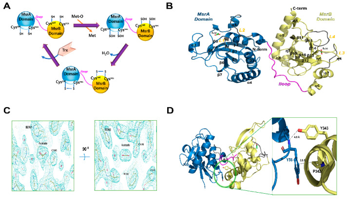
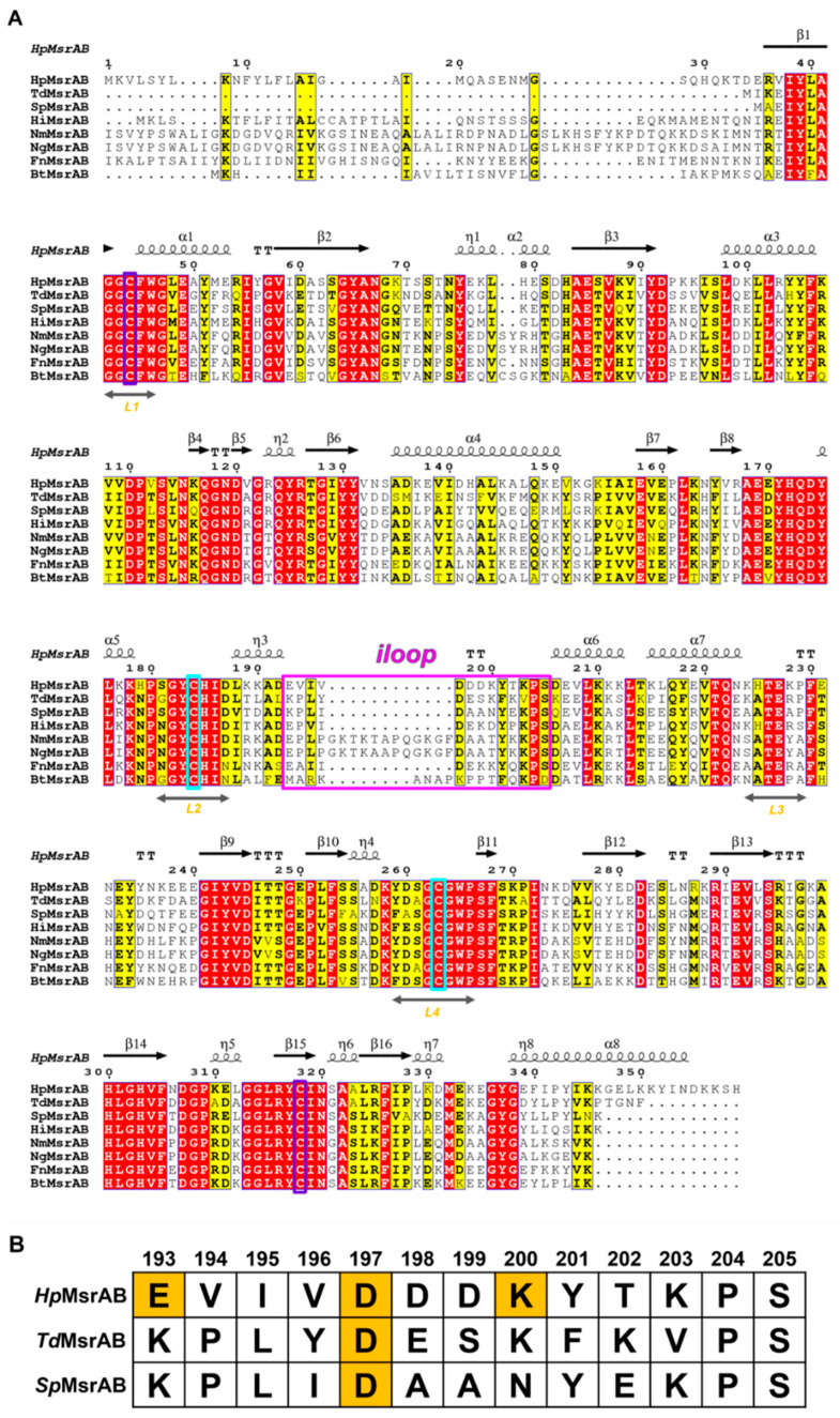
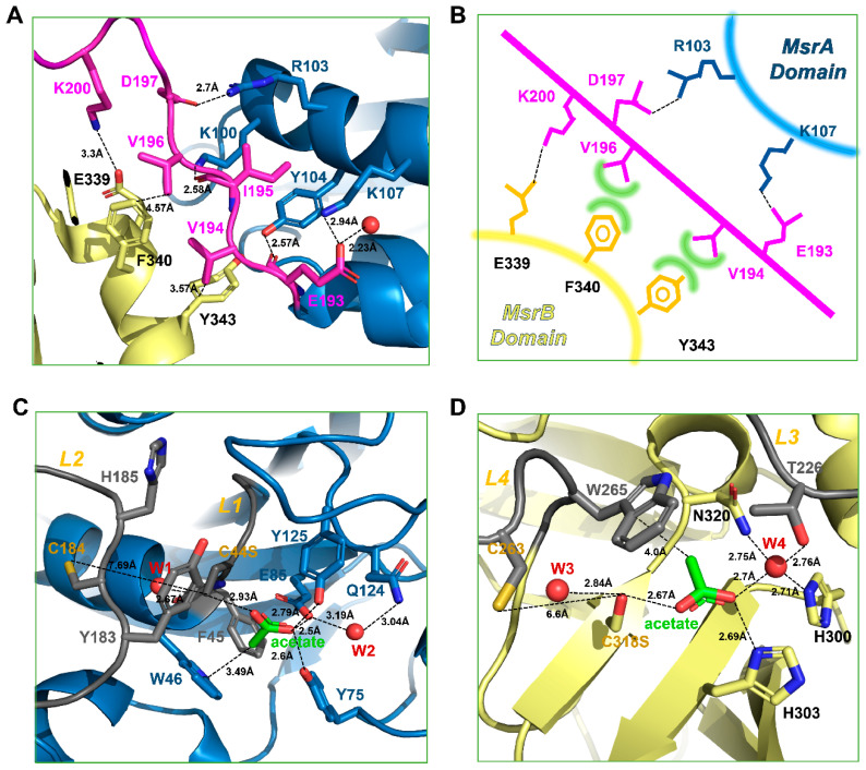
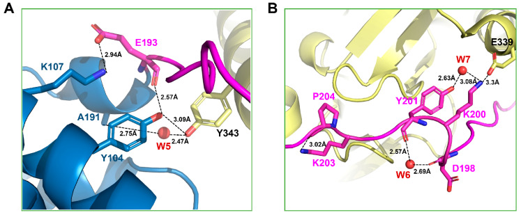
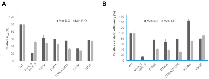
Methionine sulfoxide reductase (Msr) is a family of enzymes that reduces oxidized methionine and plays an important role in the survival of bacteria under oxidative stress conditions. MsrA and MsrB exist in a fusion protein form (MsrAB) in some pathogenic bacteria, such as Helicobacter pylori (Hp), Streptococcus pneumoniae, and Treponema denticola. To understand the fused form instead of the separated enzyme at the molecular level, we determined the crystal structure of HpMsrABC44S/C318S at 2.2 Å, which showed that a linker region (Hpiloop, 193-205) between two domains interacted with each HpMsrA or HpMsrB domain via three salt bridges (E193-K107, D197-R103, and K200-D339). Two acetate molecules in the active site pocket showed an sp2 planar electron density map in the crystal structure, which interacted with the conserved residues in fusion MsrABs from the pathogen. Biochemical and kinetic analyses revealed that Hpiloop is required to increase the catalytic efficiency of HpMsrAB. Two salt bridge mutants (D193A and E199A) were located at the entrance or tailgate of Hpiloop. Therefore, the linker region of the MsrAB fusion enzyme plays a key role in the structural stability and catalytic efficiency and provides a better understanding of why MsrAB exists in a fused form.

Keywords: MsrAB; catalytic efficiency; fusion protein; linker region.


Conflict of interest statement

The authors declare no conflict of interest.

Figures

Figure 1
Figure 1
Crystal structure of HpMsrAB. (A) Cartoon model of the general catalyzed chemical reactions of MsrAB (B) Ribbon diagram of the overall structure of HpMsrABC44S/C318S. The N-terminal domain, MsrA domain (HpMsrA, 34−192) and the C-terminal domain, MsrB domain (HpMsrB, 206−357) are colored sky blue and pale yellow, respectively. The linker region connecting HpMsrA and HpMsrB (residues 193−205), the iloop, is colored magenta. Loops (L1, 42−45 and L2, 181−187) of the HpMsrA domain and loops (L3, 224−229 and L4, 259−266) of the HpMsrB domain are colored gray. Helices (α1–α8), β-strands (β1–β16), and 310-helices (η1–η8) are labeled. Two acetate molecules are represented as stick models colored green. (C) 2Fo-Fc electron density map (1.2 sigma cutoff) around the acetate molecule in the crystal structure. (D) Close-up view of a possible hydrophobic interaction (<4 Å) between Y56 of the HpMsrA domain and P342 of the HpMsrB domain in the crystal structure. The dotted line indicates the distance between the OH of Y343 and the amide N atom of Y56 (~4.0 A), which could not form hydrogen bonds.
Figure 2
Figure 2
Multiple sequence alignment of Helicobacter pylori methionine sulfoxide reductase AB (HpMsrAB). (A) Multiple sequence alignment of MsrAB homologs in Helicobacter pylori (Hp, Swiss-Prot entry O25011), Treponema denticola (Td, Swiss-Prot entry Q73PT7), Streptococcus pneumoniae (Sp, Swiss-Prot entry P0A3Q9), Haemophilus influenzae (Hi, Swiss-Prot entry P45213), Neisseria meningitidis (Nm, Swiss-Prot entry Q9JWM8), Neisseria gonorrheae (Ng, Swiss-Prot entry P14930), Fusobacterium nucleatum (Fn, Swiss-Prot entry Q8R5 × 2), and Bacteroides thetaiotaomicron (Bt, Swiss-Prot entry Q8A4U8). Catalytic and resolving Cys residues are shown in purple and cyan, respectively. The completely conserved and similar group amino acids are represented by red and yellow letters, respectively. Hpiloop, the linker region, is displayed in a magenta box. (B) Comparison of iloops between HpMsrAB, TdMsrAB, and SpMsrAB. Partial sequence alignment of the iloops in HpMsrAB, TdMsrAB, and SpMsrAB. The residues associated with the salt bridge interactions in Hpiloops between MsrA and MsrB domains are shown in orange boxes.
Figure 3
Figure 3
Binding interface and active sites of HpMsrA and HpMsrB. (A) Zoomed view of the binding interface between HpMsrA and HpMsrB. The amino acids involved in the interaction are displayed as stick models. The residues (E193, D197, and K200) in the iloops are associated with the salt bridge interactions and the residues (E193, V194, and I195) in the iloops are associated with the hydrogen bonds or hydrophobic interactions. (B) Illustration of salt bridges and hydrophobic interactions. Dashed black lines represent salt bridges, and green curves represent hydrophobic interactions. (C) Zoomed view of the active site of the HpMsrA domain. Residues that constitute the active site are displayed as stick models. Two loops involved in forming the active site, L1 (residues 42–45) and L2 (residues 181–187), are colored gray. The acetate molecule is displayed as a stick model colored green. The water molecules are displayed as spherical models colored red. (D) Zoomed view of the active site of the HpMsrB domain. Residues that constitute the active site are displayed as stick models. Two loops associated in forming the active site, L3 (residues 224–229) and L4 (residues 259–266), are colored gray.
Figure 4
Figure 4
Interactions around the Y343 and E339 residues. (A) Magnified view of the interaction residue around Y343. The residues involved in the interaction are represented as stick models. The water molecule is represented as a spherical model colored red and (B) Close-up view of the interaction residue around E339.
Figure 5
Figure 5
Relative kinetic values. (A) Relative kcat values of HpMsrAB, HpMsrA, HpMsrB, and HpMsrAB mutants and (B) Relative catalytic efficiency (kcat/Km) of HpMsrAB, HpMsrA, HpMsrB, and HpMsrAB mutants.

Similar articles

Cited by

References

    1. Dhandayuthapani S., Jagannath C., Nino C., Saikolappan S., Sasindran S.J. Methionine sulfoxide reductase B (MsrB) of Mycobacterium smegmatis plays a limited role in resisting oxidative stress. Tuberculosis. 2009;89:S26–S32. doi: 10.1016/S1472-9792(09)70008-3. - DOI - PMC - PubMed
    1. Achilli C., Ciana A., Minetti G. The discovery of methionine sulfoxide reductase enzymes: An historical account and future perspectives. Biofactors. 2015;41:135–152. doi: 10.1002/biof.1214. - DOI - PubMed
    1. Jiang B., Moskovitz J. The functions of the mammalian methionine sulfoxide reductase system and related diseases. Antioxidants. 2018;7:122. doi: 10.3390/antiox7090122. - DOI - PMC - PubMed
    1. Kim H.-Y. The methionine sulfoxide reduction system: Selenium utilization and methionine sulfoxide reductase enzymes and their functions. Antioxid. Redox Signal. 2013;19:958–969. doi: 10.1089/ars.2012.5081. - DOI - PMC - PubMed
    1. Zhao C., Hartke A., La Sorda M., Posteraro B., Laplace J.-M., Auffray Y., Sanguinetti M. Role of methionine sulfoxide reductases A and B of Enterococcus faecalis in oxidative stress and virulence. Infect. Immun. 2010;78:3889–3897. doi: 10.1128/IAI.00165-10. - DOI - PMC - PubMed

LinkOut - more resources