. 2021 Mar 5;22(5):2618.
doi: 10.3390/ijms22052618.

Immunological Properties of Atopic Dermatitis-Associated Alopecia Areata

Affiliations

Immunological Properties of Atopic Dermatitis-Associated Alopecia Areata

Reiko Kageyama et al. Int J Mol Sci. .

Abstract

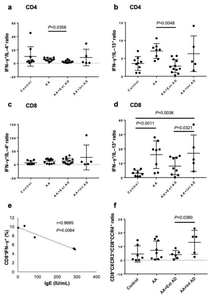
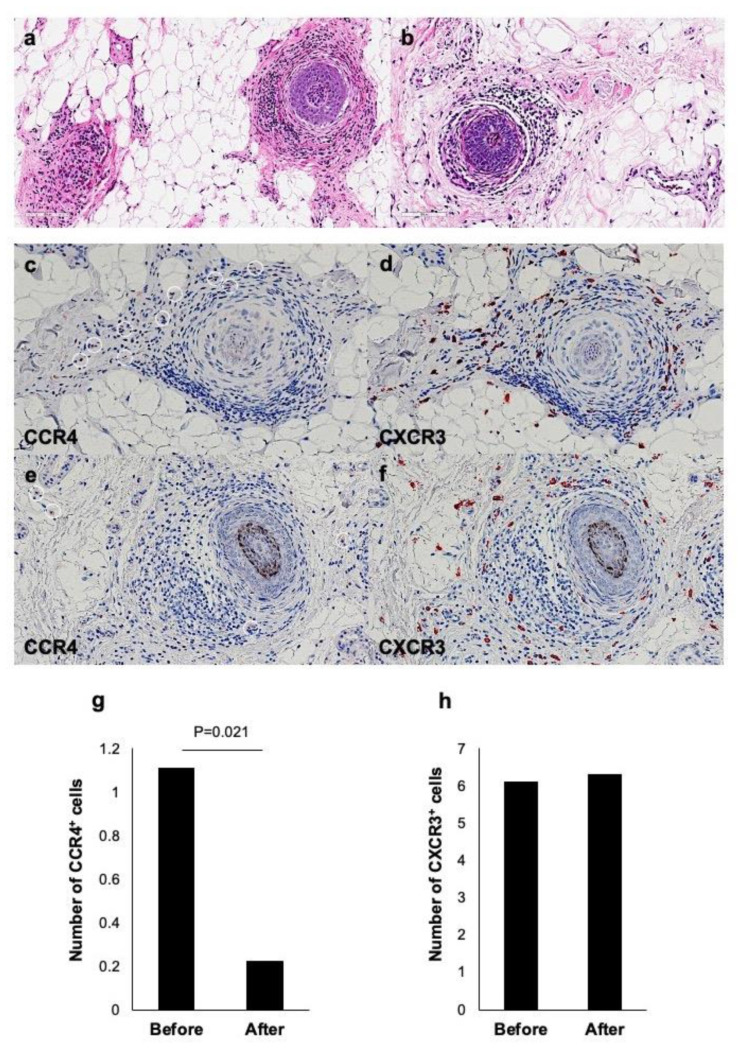
Alopecia areata (AA) is regarded as a tissue-specific and cell-mediated autoimmune disorder. Regarding the cytokine balance, AA has been considered a type 1 inflammatory disease. On the other hand, AA often complicates atopic dermatitis (AD) and AD is regarded as type 2 inflammatory disease. However, the immunological aspects of AA in relation to AD are still poorly understood. Therefore, we aim to clarify the immunological properties of AD-associated AA. In this study, we performed comparative analysis of the expression of intracytoplasmic cytokines (IFN-γ, IL-4, and IL-13), chemokine receptors (CXCR3 and CCR4) in peripheral blood which were taken from healthy controls, non-atopic AA patients, AA patients with extrinsic AD, and AA patients with intrinsic AD by flowcytometric analysis. We also compared the scalp skin samples taken from AA patients with extrinsic AD before and after treatment with dupilumab. In non-atopic AA patients, the ratios of CD4+IFN-γ+ cells to CD4+IL-4+ cells and CD4+IFN-γ+ cells to CD4+IL-13+ cells were higher than those in AA patients with extrinsic AD. Meanwhile, the ratio of CD8+IFN-γ+ cells to CD8+IL-13+ cells was significantly higher in the non-atopic AA than in the healthy controls. In AA patients with extrinsic AD, the skin AA lesion showed dense infiltration of not only CXCR3+ cells but also CCR4+ cells around hair bulb before dupilumab treatment. However, after the treatment, the number of CXCR3+ cells had no remarkable change while the number of CCR4+ cells significantly decreased. These results indicate that the immunological condition of AA may be different between atopic and non-atopic patients and between extrinsic and intrinsic AD patients. Our study provides an important notion that type 2 immunity may participate in the development of AA in extrinsic AD patients. It may be considered that the immunological state of non-atopic AA is different from that of atopic AA.

Keywords: IFN-γ; IL-13; alopecia areata; extrinsic atopic dermatitis; intrinsic atopic dermatitis.


Conflict of interest statement

The authors declare no conflict of interest.

Figures

Figure 1
Figure 1
Ratio of Th1/Tc1 to Th2/Tc2 in peripheral blood of non-atopic and atopic patients with AA. (ad) The expression of cytoplasmic cytokines was analyzed by flow cytometric analysis. The ratios of IFN–γ/IL-4 and IFN–γ/IL-13 in CD4+ (a,b) and CD8+ T cells (c,d) were measured in normal control subjects, non-atopic AA, and AA with extrinsic or intrinsic AA. (e) The correlation between the frequency of IFN–γ-producing cells and the serum IgE level was analyzed in the patients with non-atopic AA. (f) The ratio of CD8+CXCR3+ (Tc1) to CD8+CCR4+ (Tc2) cells was analyzed by flow cytometric analysis in normal control subjects, non-atopic AA, and AA with extrinsic or intrinsic AA.
Figure 2
Figure 2
Significant correlation between SALT and EASI score in the patients with AA and extrinsic AD. The correlation between SALT and EASI score were analyzed in the twelve AA patients with extrinsic AD. Interestingly, the two scores were found to be significantly correlated by Spearman’s correlation coefficient.
Figure 3
Figure 3
Clinical improvement of AD and AA by dupilumab treatment. (ad) Representative case of AA with extrinsic AD before and 6 months after dupilumab therapy. (e,f) Changes of EASI scores (e) and SALT scores (f) in 6 patients with extrinsic AD and AA before and 6 months after dupilumab therapy. (gj) Representative case of AA with extrinsic AD before and 6 months after dupilumab therapy. (k) The CXCR3/CCR4 ratio in the AA patients with extrinsic AD.
Figure 4
Figure 4
Histological and immunohistochemical findings in a case of AA with extrinsic AD (case 2). H.E. staining before (a) and 16 weeks after (b) dupilumab therapy. Immunostaining of CCR4 and CXCR3 before (c,d) and 16 weeks after (e,f) dupilumab therapy. (g,h) The number of CCR4 negative and CXCR3 positive cells before and 16 weeks after dupilumab therapy.

References

    1. Ito T., Ito N., Bettermann A., Tokura Y., Takigawa M., Paus R. Collapse and resto-ration of MHC class-I-dependent immune privilege: Exploiting the human hair follicle as a model. Am. J. Pathol. 2004;164:623–634. doi: 10.1016/S0002-9440(10)63151-3. - DOI - PMC - PubMed
    1. Paus R., Ito N., Takigawa M., Ito T. The hair follicle and immune privilege. J. Investig. Dermatol. Symp. Proc. 2003;8:188–194. doi: 10.1046/j.1087-0024.2003.00807.x. - DOI - PubMed
    1. Żeberkiewicz M., Rudnicka L., Malejczyk J. Immunology of alopecia areata. Cent. Eur. J. Immunol. 2020;45:325–333. doi: 10.5114/ceji.2020.101264. - DOI - PMC - PubMed
    1. McElwe K.J., Gilhar A., Tobin D.J., Ramot Y., Sundberg J.P., Nakamura M., Bertolini M., Inui S., Tokura Y., King L.E., Jr., et al. What causes alopecia areata? Exp. Dermatol. 2013;22:609–626. doi: 10.1111/exd.12209. - DOI - PMC - PubMed
    1. Gilhar A., Kam Y., Assy B., Kalish R.S. Alopecia areata induced in C3H/HeJ mice by interferon-gamma: Evidence for loss of immune privilege. J. Invest. Dermatol. 2005;124:288–289. doi: 10.1111/j.0022-202X.2004.23580.x. - DOI - PubMed

MeSH terms

LinkOut - more resources