Overview of Neo-Vascular Lesions after Delivery or Miscarriage
- PMID: 33807766
- PMCID: PMC7961487
- DOI: 10.3390/jcm10051084
Overview of Neo-Vascular Lesions after Delivery or Miscarriage
Abstract
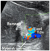
The concept of intrauterine neo-vascular lesions after pregnancy, initially called placental polyps, has changed gradually. Now, based on diagnostic imaging, such lesions are defined as retained products of conception (RPOC) with vascularization. The lesions appear after delivery or miscarriage, and they are accompanied by frequent abundant vascularization in the myometrium attached to the remnant. Many of these vascular lesions have been reported to resolve spontaneously within a few months. Acquired arteriovenous malformations (AVMs) must be considered in the differential diagnosis of RPOC with vascularization. AVMs are errors of morphogenesis. The lesions start to be constructed at the time of placenta formation. These lesions do not show spontaneous regression. Although these two lesions are recognized as neo-vascular lesions, neo-vascular lesions on imaging may represent conditions other than these two lesions (e.g., peritrophoblastic flow, uterine artery pseudoaneurysm, and villous-derived malignancies). Detecting vasculature at the placenta-myometrium interface and classifying vascular diseases according to hemodynamics in the remnant would facilitate the development of specific treatments.
Keywords: arteriovenous malformations; neo-vascular lesion; placental polyps; retained products of conception.
Conflict of interest statement
The authors declare that they have no conflicts of interest.
Figures


References
-
- Baer B.F. Placental Polypus which simulated malignant disease of the uterus. Phila. Med. Times. 1884;15:175.
-
- Hagstrom H.T. Late puerperal hemorrhages due to placental polyp. Am. J. Obstet. Gynecol. 1940;39:879–881. doi: 10.1016/S0002-9378(40)91059-6. - DOI
-
- Hoberman L.K., Hawkinson J.A., Beecham C.T. Placental polyps: Report of three cases. Obstet. Gynecol. 1963;22:25–29. - PubMed
-
- Swan R.W., Woodruff J.D. Retained products of conception: Histologic viability of placental polyps. Obstet. Gynecol. 1969;34:506–514. - PubMed
Publication types
LinkOut - more resources
Full Text Sources
Other Literature Sources

