. 2021 Mar 5;10(3):542.
doi: 10.3390/foods10030542.

Analysis of Biofilm Formation on the Surface of Organic Mung Bean Seeds, Sprouts and in the Germination Environment

Affiliations

Analysis of Biofilm Formation on the Surface of Organic Mung Bean Seeds, Sprouts and in the Germination Environment

Marcin Kruk et al. Foods. .

Abstract

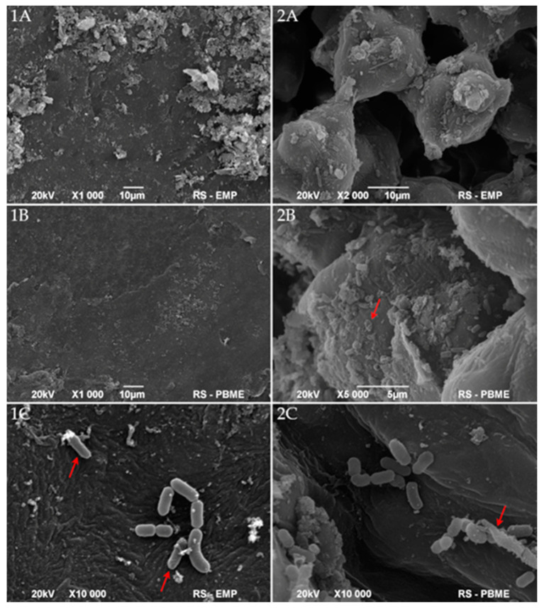
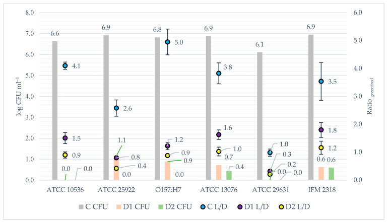
This study aimed to analyse the impact of sanitation methods on the formation of bacterial biofilms after disinfection and during the germination process of mung bean on seeds and in the germination environment. Moreover, the influence of Lactobacillus plantarum 299v on the growth of the tested pathogenic bacteria was evaluated. Three strains of Salmonella and E. coli were used for the study. The colony forming units (CFU), the crystal violet (CV), the LIVE/DEAD and the gram fluorescent staining, the light and the scanning electron microscopy (SEM) methods were used. The tested microorganisms survive in a small number. During germination after disinfection D2 (20 min H2O at 60 °C, then 15 min in a disinfecting mixture consisting of H2O, H2O2 and CH₃COOH), the biofilms grew most after day 2, but with the DP2 method (D2 + L. plantarum 299v during germination) after the fourth day. Depending on the method used, the second or fourth day could be a time for the introduction of an additional growth-limiting factor. Moreover, despite the use of seed disinfection, their germination environment could be favourable for the development of bacteria and, consequently, the formation of biofilms. The appropriate combination of seed disinfection methods and growth inhibition methods at the germination stage will lead to the complete elimination of the development of unwanted microflora and their biofilms.

Keywords: biofilm; mung bean; organic food; pathogen; probiotic; sprouts.


Conflict of interest statement

The authors declare no conflict of interest.

Figures

Figure 1
Figure 1
Schematic diagram of the experiment and disinfection methods (D1, D2, DP1, DP2); probiotic—L. plantarum 299v. D1—disinfected with the 1st method; D2—disinfected with the 2nd method; DP1—disinfected with the 1st method + L. plantarum 299v; DP2—disinfected with the 2nd method + L. plantarum 299v; SEM—Scanning Electron Microscopy; CFU—Colony Formation Units; CV—Crystal Violet method.
Figure 2
Figure 2
Scanning electron micrographs of whole native bean (A), hilum (C), and coat (B,D) of mung bean.
Figure 3
Figure 3
Scanning electron micrographs of coats (1) and hila (2) of mung bean inoculated with pathogens (A(1,2)) and after then disinfected with D2 method (B(1,2)) or DP2 method (C(1,2)); arrows indicate destroyed (2(B,C)) or damaged (1C) cells.
Figure 4
Figure 4
Formation of biofilms by E. coli ATCC 10536 (A); E. coli O157:H7 (B); E.coli ATCC 25922 (C); S. enteritadis ATCC 13076 (D); S. enteritadis ATCC 29631 (E); S. hofit IFM 2318 (F) on the surface of 24-well microplate for 6 days at 22 °C. C—control, not disinfected with any method; D1—disinfected with the 1st method; D2—disinfected with the 2nd method; DP1—disinfected with the 1st method + L. plantarum 299v; DP2—disinfected with the 2nd method + L. plantarum 299v; C —; D1—; D2—; DP1—; DP2—; n = 4.
Figure 5
Figure 5
Light microscopy micrographs of biofilms formed during mung bean germination. Explanations: The numbers (1,2,3,4,5,6) refer to the strain. (1)—E. coli ATCC 10536; (2)—E.coli ATCC 25922; (3)—E. coli O157:H7; (4)—S. enteritadis ATCC 13076; (5)—S. enteritadis ATCC 29631; (6)—S. hofit IFM 2318. The letters (a,b,c,d) refer to the disinfection method. (a)—D1 disinfected with the 1st method; (b)—D2 disinfected with the 2nd method; (c)—DP1 disinfected with the 1st method + L. plantarum 299v; (d)—DP2 disinfected with the 2nd method + L. plantarum 299v; Bacteria were observed at 1000× magnification.
Figure 6
Figure 6
The survival of tested strains after disinfection procedure measured by fluorescence staining vs. CFU analysis. Explanations: L/D—LIVE/DEAD method = fluorescence staining; CFU—colony forming units method; bars refer to CFU results; dots refer to fluorescence staining analysis; C—control, not disinfected with any method; D1—disinfected with the 1st method; D2—disinfected with the 2nd method; DP1—disinfected with the 1st method + L. plantarum 299v; DP2—disinfected with the 2nd method + L. plantarum 299v.

Similar articles

Cited by

References

    1. Benincasa P., Falcinelli B., Lutts S., Stagnari F., Galieni A. Sprouted Grains: A Comprehensive Review. Nutrients. 2019;11:421. doi: 10.3390/nu11020421. - DOI - PMC - PubMed
    1. Iacumin L., Comi G. Microbial Quality of Raw and Ready-to-Eat Mung Bean Sprouts Produced in Italy. Food Microbiol. 2019;82:371–377. doi: 10.1016/j.fm.2019.03.014. - DOI - PubMed
    1. Mustar S., Nazaimoon W.M.W. The Effect of Sanitizers on the Native Microflora of Mung Bean Seeds (Vigna Radiata) J. Food Technol. 2010;8:234–238. doi: 10.3923/jftech.2010.234.238. - DOI
    1. Gabriel A.A. Microbial Quality of Chlorine Soaked Mung Bean Seeds and Sprouts. Food Sci. Technol. Res. 2005;11:95–100. doi: 10.3136/fstr.11.95. - DOI
    1. Zhang C., Lu Z., Li Y., Shang Y., Zhang G., Cao W. Reduction of Escherichia Coli O157:H7 and Salmonella Enteritidis on Mung Bean Seeds and Sprouts by Slightly Acidic Electrolyzed Water. Food Control. 2011;22:792–796. doi: 10.1016/j.foodcont.2010.11.018. - DOI

LinkOut - more resources