

From Mitochondria to Atherosclerosis: The Inflammation Path
- PMID: 33807807
- PMCID: PMC8000234
- DOI: 10.3390/biomedicines9030258
From Mitochondria to Atherosclerosis: The Inflammation Path
Abstract
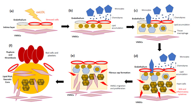
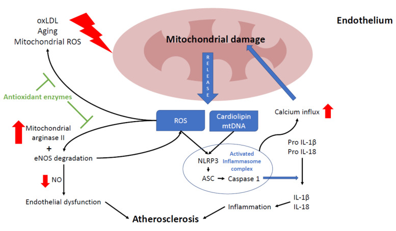
Inflammation is a key process in metazoan organisms due to its relevance for innate defense against infections and tissue damage. However, inflammation is also implicated in pathological processes such as atherosclerosis. Atherosclerosis is a chronic inflammatory disease of the arterial wall where unstable atherosclerotic plaque rupture causing platelet aggregation and thrombosis may compromise the arterial lumen, leading to acute or chronic ischemic syndromes. In this review, we will focus on the role of mitochondria in atherosclerosis while keeping inflammation as a link. Mitochondria are the main source of cellular energy. Under stress, mitochondria are also capable of controlling inflammation through the production of reactive oxygen species (ROS) and the release of mitochondrial components, such as mitochondrial DNA (mtDNA), into the cytoplasm or into the extracellular matrix, where they act as danger signals when recognized by innate immune receptors. Primary or secondary mitochondrial dysfunctions are associated with the initiation and progression of atherosclerosis by elevating the production of ROS, altering mitochondrial dynamics and energy supply, as well as promoting inflammation. Knowing and understanding the pathways behind mitochondrial-based inflammation in atheroma progression is essential to discovering alternative or complementary treatments.
Keywords: NLRP3; atherosclerosis; inflammasome; inflammation; mitochondria; reactive oxygen species.
Conflict of interest statement
The authors declare that the research was conducted in the absence of any commercial or financial relationships that could be construed as a potential conflict of interest.
Figures
Similar articles
-
Mitochondrial Dysfunction in Vascular Wall Cells and Its Role in Atherosclerosis.Int J Mol Sci. 2021 Aug 20;22(16):8990. doi: 10.3390/ijms22168990. Int J Mol Sci. 2021. PMID: 34445694 Free PMC article. Review.
-
Do Mitochondrial DNA Mutations Play a Key Role in the Chronification of Sterile Inflammation? Special Focus on Atherosclerosis.Curr Pharm Des. 2021;27(2):276-292. doi: 10.2174/1381612826666201012164330. Curr Pharm Des. 2021. PMID: 33045961 Review.
-
Mitochondrial Dysfunction: The Hidden Player in the Pathogenesis of Atherosclerosis?Int J Mol Sci. 2023 Jan 6;24(2):1086. doi: 10.3390/ijms24021086. Int J Mol Sci. 2023. PMID: 36674602 Free PMC article. Review.
-
Melatonin Ameliorates the Progression of Atherosclerosis via Mitophagy Activation and NLRP3 Inflammasome Inhibition.Oxid Med Cell Longev. 2018 Sep 4;2018:9286458. doi: 10.1155/2018/9286458. eCollection 2018. Oxid Med Cell Longev. 2018. PMID: 30254716 Free PMC article.
-
Ambiguities in NLRP3 inflammasome regulation: is there a role for mitochondria?Biochim Biophys Acta. 2014 Apr;1840(4):1433-40. doi: 10.1016/j.bbagen.2013.08.014. Epub 2013 Aug 27. Biochim Biophys Acta. 2014. PMID: 23994495 Review.
Cited by
-
Lipids and Lipoproteins in Health and Disease.Biomedicines. 2021 Dec 31;10(1):87. doi: 10.3390/biomedicines10010087. Biomedicines. 2021. PMID: 35052767 Free PMC article.
-
A review on current advancement in zebrafish models to study chronic inflammatory diseases and their therapeutic targets.Heliyon. 2024 May 23;10(11):e31862. doi: 10.1016/j.heliyon.2024.e31862. eCollection 2024 Jun 15. Heliyon. 2024. PMID: 38867970 Free PMC article. Review.
-
Mitochondrial Dysfunction in Endothelial Progenitor Cells: Unraveling Insights from Vascular Endothelial Cells.Biology (Basel). 2024 Jan 23;13(2):70. doi: 10.3390/biology13020070. Biology (Basel). 2024. PMID: 38392289 Free PMC article. Review.
-
The two coin sides of bacterial extracellular membrane nanovesicles: atherosclerosis trigger or remedy.Discov Nano. 2024 Nov 12;19(1):179. doi: 10.1186/s11671-024-04149-8. Discov Nano. 2024. PMID: 39532781 Free PMC article. Review.
-
The role of systemic immune-inflammatory index in predicting contrast-induced nephropathy in non-ST-segment elevation myocardial infarction cases.Biomark Med. 2024;18(21-22):937-944. doi: 10.1080/17520363.2024.2415284. Epub 2024 Oct 29. Biomark Med. 2024. PMID: 39469834
References
Publication types
Grants and funding
LinkOut - more resources
Full Text Sources
Other Literature Sources
