

Neuroimaging of Acute Intracerebral Hemorrhage
- PMID: 33807843
- PMCID: PMC7962049
- DOI: 10.3390/jcm10051086
Neuroimaging of Acute Intracerebral Hemorrhage
Abstract
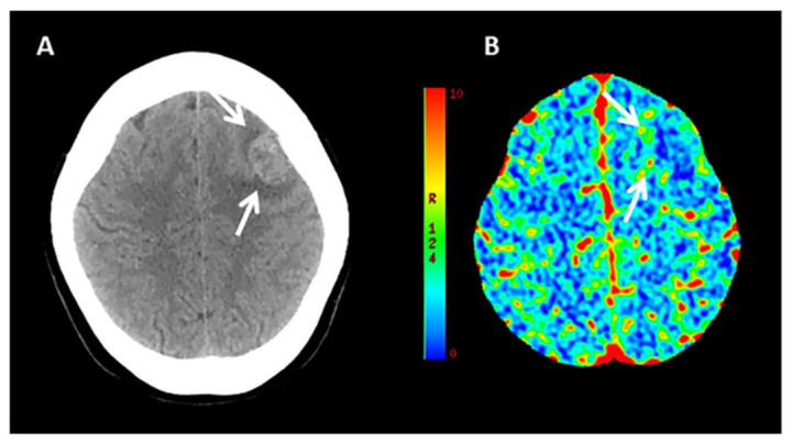
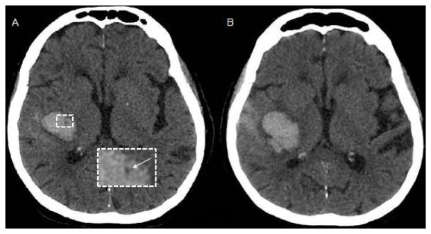
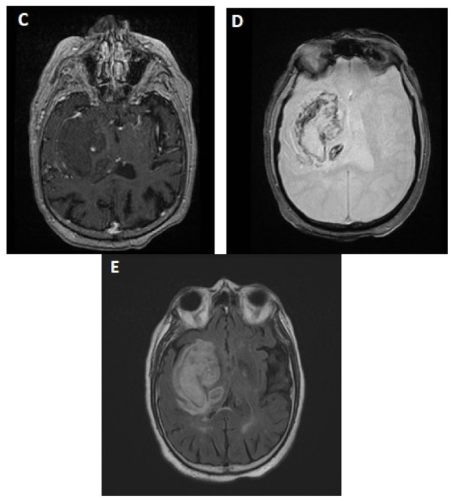
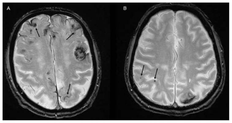
Intracerebral hemorrhage (ICH) accounts for 10% to 20% of all strokes worldwide and is associated with high morbidity and mortality. Neuroimaging is clinically important for the rapid diagnosis of ICH and underlying etiologies, but also for identification of ICH expansion, often as-sociated with an increased risk for poor outcome. In this context, rapid assessment of early hema-toma expansion risk is both an opportunity for therapeutic intervention and a potential hazard for hematoma evacuation surgery. In this review, we provide an overview of the current literature surrounding the use of multimodal neuroimaging of ICH for etiological diagnosis, prediction of early hematoma expansion, and prognostication of neurological outcome. Specifically, we discuss standard imaging using computed tomography, the value of different vascular imaging modalities to identify underlying causes and present recent advances in magnetic resonance imaging and computed tomography perfusion.
Keywords: ICH expansion; NCCT markers; imaging; intracerebral hemorrhage; outcome; spot sign.
Conflict of interest statement
The authors declare no conflict of interest.
Figures
References
-
- Johnson C.O., Nguyen M., Roth G.A., Nichols E., Alam T., Abate D., Abd-Allah F., Abdelalim A., Abraha H.N., Abu-Rmeileh N.M., et al. Global, regional, and national burden of stroke, 1990–2016: A systematic analysis for the Global Burden of Disease Study 2016. Lancet Neurol. 2019;18:357–375. doi: 10.1016/S1474-4422(19)30034-1. - DOI - PMC - PubMed
-
- Rodrigues M.A., Samarasekera N., Lerpiniere C., Humphreys C., McCarron M.O., White P.M., Nicoll J.A.R., Sudlow C.L.M., Cordonnier C., Wardlaw J.M., et al. The Edinburgh CT and genetic diagnostic criteria for lobar intracerebral haemorrhage associated with cerebral amyloid angiopathy: Model development and diagnostic test accuracy study. Lancet Neurol. 2018;17:232–240. doi: 10.1016/S1474-4422(18)30006-1. - DOI - PMC - PubMed
Publication types
LinkOut - more resources
Full Text Sources
Other Literature Sources
