Review
. 2021 Mar 5;11(3):387.
doi: 10.3390/biom11030387.

Intracellular Toxic AGEs (TAGE) Triggers Numerous Types of Cell Damage

Affiliations
Review

Intracellular Toxic AGEs (TAGE) Triggers Numerous Types of Cell Damage

Masayoshi Takeuchi et al. Biomolecules. .

Abstract

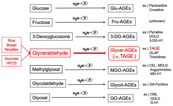
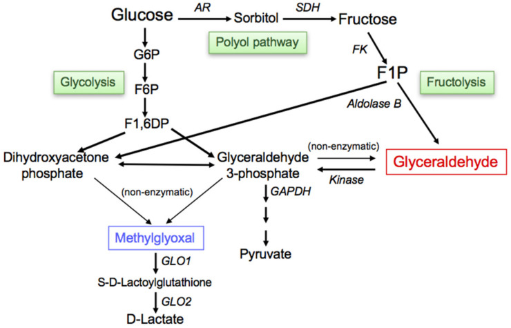
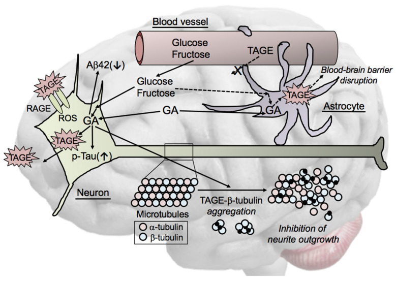
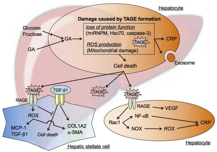
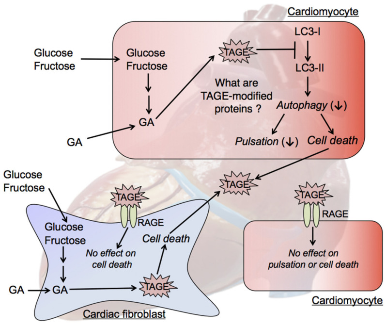
The habitual intake of large amounts of sugar, which has been implicated in the onset/progression of lifestyle-related diseases (LSRD), induces the excessive production of glyceraldehyde (GA), an intermediate of sugar metabolism, in neuronal cells, hepatocytes, and cardiomyocytes. Reactions between GA and intracellular proteins produce toxic advanced glycation end-products (toxic AGEs, TAGE), the accumulation of which contributes to various diseases, such as Alzheimer's disease, non-alcoholic steatohepatitis, and cardiovascular disease. The cellular leakage of TAGE affects the surrounding cells via the receptor for AGEs (RAGE), thereby promoting the onset/progression of LSRD. We demonstrated that the intracellular accumulation of TAGE triggered numerous cellular disorders, and also that TAGE leaked into the extracellular space, thereby increasing extracellular TAGE levels in circulating fluids. Intracellular signaling and the production of reactive oxygen species are affected by extracellular TAGE and RAGE interactions, which, in turn, facilitate the intracellular generation of TAGE, all of which may contribute to the pathological changes observed in LSRD. In this review, we discuss the relationships between intracellular TAGE levels and numerous types of cell damage. The novel concept of the "TAGE theory" is expected to open new perspectives for research into LSRD.

Keywords: Alzheimer’s disease (AD); advanced glycation end-products (AGEs); cardiovascular disease (CVD); lifestyle-related diseases (LSRD); non-alcoholic steatohepatitis (NASH); toxic AGEs (TAGE).


Conflict of interest statement

The authors declare that no conflict of interest exist.

Figures

Figure 1
Figure 1
Various routes for the in vivo production of advanced glycation end-products (AGEs). Reducing sugars, including glucose, fructose, and glyceraldehyde, react non-enzymatically with the amino/guanidino groups of proteins, resulting in the formation of reversible Schiff bases and Amadori products/Heyns products. Further complex reactions involving these early glycation products, such as rearrangement, dehydration, and condensation reactions, lead to irreversibly cross-linked, heterogeneous fluorescent derivatives, named AGEs. Glu-AGEs: glucose-derived AGEs; Fru-AGEs: fructose-derived AGEs; 3-DG-AGEs: 3-deoxyglucosone-derived AGEs; Glycer-AGEs: glyceraldehyde-derived AGEs; TAGE: toxic AGEs; MGO-AGEs: methylglyoxal-derived AGEs; Glycol-AGEs: glycolaldehyde-derived AGEs; GO-AGEs: glyoxal-derived AGEs; DOLD: 3-deoxyglucosone-lysine dimer; 3-DG-H1: 3-deoxyglucosone-derived hydroimidazolone; GLAP: glyceraldehyde-derived pyridinium; CEL: Nε-(carboxyethyl)lysine; MOLD: methylglyoxal-lysine dimer; MG-H1: methylglyoxal-derived hydroimidazolone 1; GA-pyridine: glycolaldehyde-derived pyridine; CML: Nε-(carboxymethyl)lysine; GOLD: glyoxal-lysine dimer; G-H1; glyoxal-derived hydroimidazolone 1; HFCS: high-fructose corn syrup; P-NH2: free amino residue of a protein.
Figure 2
Figure 2
Routes for the in vivo production of glyceraldehyde (GA)/methylglyoxal (MGO). The glycolytic intermediate glyceraldehyde 3-phosphate (GA3P) is generally catabolized (glycolysis) by the enzyme GA3P dehydrogenase (GAPDH). However, decreases in the enzymatic activity of GAPDH result in the intracellular accumulation of GA3P. As a consequence, GA3P is metabolized via an alternative pathway, which increases the concentration of GA. Fructokinase phosphorylates fructose to fructose 1-phosphate (F1P), which is then converted into dihydroxyacetone phosphate (DHAP) and GA by aldolase B (fructolysis). MGO is mainly produced as a byproduct of non-enzymatic reactions with GA3P or DHAP during glycolysis. The most effective MGO metabolic pathway is the glyoxalase system, which converts MGO to D-lactate. G6P: glucose 6-phosphate; F6P: fructose 6-phosphate; F1,6DP: fructose 1,6-diphosphate; AR: aldose reductase; SDH: sorbitol dehydrogenase; FK: fructokinase; GLO1: glyoxalase 1; GLO2: glyoxalase 2.
Figure 3
Figure 3
Cytotoxicity of TAGE in neuronal cells. TAGE mainly localize to neuronal cell bodies of neurons in AD brains. GA induces the generation of TAGE and exhibits cytotoxicity towards neuronal cells. Neuronal cell death may induce the extracellular leakage of TAGE, which affects the surrounding cells via the TAGE-RAGE-ROS system. TAGE reduce the concentration of amyloid β1-42 (Aβ42) in culture media and increase tau phosphorylation. β-Tubulin is a target of TAGE-modified proteins. GA induces abnormal β-tubulin aggregation and inhibits neurite outgrowth through the formation of TAGE-β-tubulin. We also hypothesize that TAGE-targeted proteins are involved in GA-induced blood-brain barrier disruption in astrocytes. GA: glyceraldehyde; RAGE: receptor for AGEs; ROS: reactive oxygen species; TAGE: toxic AGEs.
Figure 4
Figure 4
TAGE cytotoxicity against hepatocytes and hepatic stellate cells (HSC). The chronic intake of excessive amounts of sugar-sweetened beverages and processed food products increases the concentration of the sugar metabolite GA in hepatocytes. GA accumulation in these cells promotes TAGE modifications in cellular components. Loss of protein function and mitochondrial membrane abnormalities are associated with the formation of TAGE. ROS are also produced intracellularly and ultimately cause hepatocyte death. Hepatocyte death may cause TAGE-modified proteins to leak from cells, and extracellular TAGE affect the surrounding cells via the TAGE-RAGE axis. The TAGE-RAGE axis exerts inflammatory effects within hepatocytes. HSC are also affected by extracellular TAGE. ROS production and fibrosis are promoted in HSC through the activation of TAGE-RAGE signaling. In addition, TAGE inhibit TGF-β1-induced cell death and help to maintain the number of HSC, resulting in increased extracellular matrix molecule production and, ultimately, fibrosis. GA: glyceraldehyde; TAGE: toxic AGEs; hnRNPM: heterogenous nuclear ribonucleoprotein M; Hsc70: heat shock cognate 70; ROS: reactive oxygen species; CRP: C-reactive protein; RAGE: receptor for AGEs; MCP-1: monocyte chemoattractant protein-1; TGF-β1: transforming growth factor-β1; COL1A2: collagen-type Iα2 chain; α-SMA: α-smooth muscle actin; VEGF: vascular endothelial growth factor; Rac1: Ras-related C3 botulinum toxin substrate 1; NF-κB: nuclear factor kappa B; NOX: nicotinamide adenine dinucleotide phosphate reduced (NADPH) oxidase.
Figure 5
Figure 5
TAGE cytotoxicity against cardiomyocytes and cardiac fibroblasts. GA accumulation in cardiomyocytes leads to TAGE-induced modifications in cellular components, including proteins. TAGE may be released from dead cardiomyocytes; however, extracellular TAGE do not suppress cardiomyocyte pulsation or induce cell death in cardiomyocytes via the TAGE-RAGE axis. The intracellular generation of TAGE in cardiac fibroblasts and their cytotoxicity, and the effects of extracellular TAGE on cardiac fibroblasts have not yet been elucidated. GA: glyceraldehyde; LC3 (MAP1LC3): microtubule-associated protein light chain 3; RAGE: receptor for AGEs; TAGE: toxic AGEs.

References

    1. Saeedi P., Petersohn I., Salpea P., Malanda B., Karuranga S., Unwin N., Colagiuri S., Guariguata L., Motala A.A., Ogurtsova K., et al. Global and regional diabetes prevalence estimates for 2019 and projections for 2030 and 2045: Results from the International Diabetes Federation Diabetes Atlas, 9th edition. Diabetes Res. Clin. Pract. 2019;157:107843. doi: 10.1016/j.diabres.2019.107843. - DOI - PubMed
    1. Wang M., Yu M., Fang L., Hu R.Y. Association between sugar-sweetened beverages and type 2 diabetes: A meta-analysis. J. Diabetes Investig. 2015;6:360–366. doi: 10.1111/jdi.12309. - DOI - PMC - PubMed
    1. Imamura F., O’Connor L., Ye Z., Mursu J., Hayashino Y., Bhupathiraju S.N., Forouhi N.G. Consumption of sugar sweetened beverages, artificially sweetened beverages, and fruit juice and incidence of type 2 diabetes: Systematic review, meta-analysis, and estimation of population attributable fraction. Br. J. Sports Med. 2016;50:496–504. doi: 10.1136/bjsports-2016-h3576rep. - DOI - PMC - PubMed
    1. Malik V.S. Sugar sweetened beverages and cardiometabolic health. Curr. Opin. Cardiol. 2017;32:572–579. doi: 10.1097/HCO.0000000000000439. - DOI - PubMed
    1. Schwingshackl L., Hoffmann G., Lampousi A.M., Knüppel S., Iqbal K., Schwedhelm C., Bechthold A., Schlesinger S., Boeing H. Food groups and risk of type 2 diabetes mellitus: A systematic review and meta-analysis of prospective studies. Eur. J. Epidemiol. 2017;32:363–375. doi: 10.1007/s10654-017-0246-y. - DOI - PMC - PubMed

Publication types

MeSH terms

Substances

LinkOut - more resources