Review
. 2021 Mar 30;10(4):754.
doi: 10.3390/cells10040754.

Molecular Chaperones in Osteosarcoma: Diagnosis and Therapeutic Issues

Affiliations
Review

Molecular Chaperones in Osteosarcoma: Diagnosis and Therapeutic Issues

Morgane Lallier et al. Cells. .

Abstract

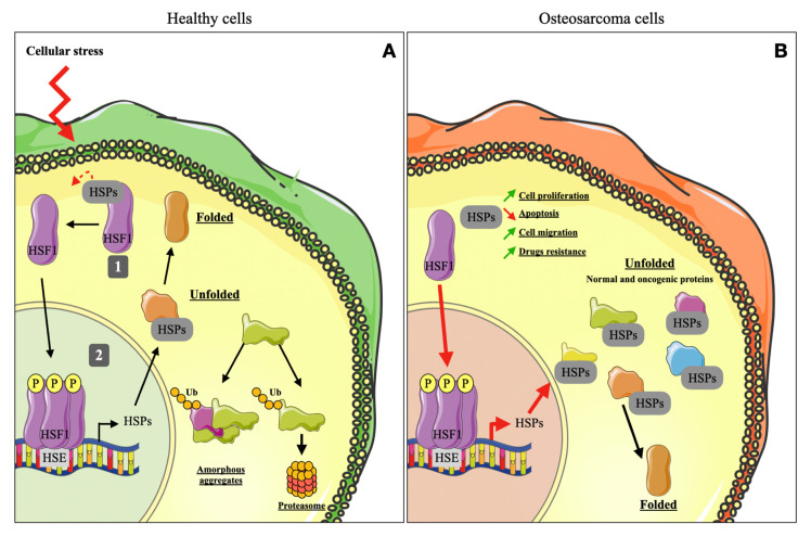
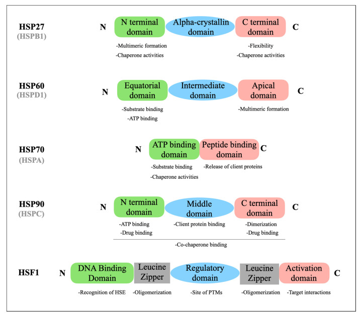
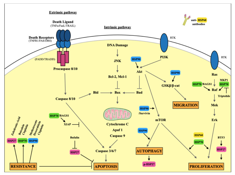
Osteosarcoma (OS) is the most common form of primary bone tumor affecting mainly children and young adults. Despite therapeutic progress, the 5-year survival rate is 70%, but it drops drastically to 30% for poor responders to therapies or for patients with metastases. Identifying new therapeutic targets is thus essential. Heat Shock Proteins (HSPs) are the main effectors of Heat Shock Response (HSR), the expression of which is induced by stressors. HSPs are a large family of proteins involved in the folding and maturation of other proteins in order to maintain proteostasis. HSP overexpression is observed in many cancers, including breast, prostate, colorectal, lung, and ovarian, as well as OS. In this article we reviewed the significant role played by HSPs in molecular mechanisms leading to OS development and progression. HSPs are directly involved in OS cell proliferation, apoptosis inhibition, migration, and drug resistance. We focused on HSP27, HSP60, HSP70 and HSP90 and summarized their potential clinical uses in OS as either biomarkers for diagnosis or therapeutic targets. Finally, based on different types of cancer, we consider the advantage of targeting heat shock factor 1 (HSF1), the major transcriptional regulator of HSPs in OS.

Keywords: HSF1; HSPs; bone tumor; osteosarcoma.


Conflict of interest statement

The authors declare no conflict of interest.

Figures

Figure 1
Figure 1
HSR and HSP functions in a healthy cell and OS cell. (A) Under physiological conditions, HSF1 is complexed with HSPs and is located in the cell cytoplasm (1). During cellular stress, HSF1 is phosphorylated, trimerized and translocated into the nucleus where it binds to the HSE motif and initiates transcription of HSPs (2). Some unfolded proteins are taken over by HSPs to be refolded. Others are ubiquitinylated and degraded by the proteasome. Accumulation of unfolded proteins leads to amorphous aggregations, compromising the cell’s survival. (B) In a cancer cell, HSF1 is over-activated and initiates the transcription of numerous HSPs, which take over normal and oncogenic unfolded proteins and refold them, thereby averting proteomic instability. Furthermore, HSPs are directly involved in different mechanisms that promote cell survival, such as cell proliferation and migration, apoptosis and drug resistance. HSE: Heat Shock Element; HSF1: Heat Shock Factor 1; HSPs: Heat Shock Proteins; Ub: Ubiquitin; P: Phosphorylation.
Figure 2
Figure 2
Schematic structure of HSP27 (HSPB1), HSP60 (HSPD1), HSP70 (HSPA), HSP90 (HSPC) and HSF1. The main functions of each domain are specified. HSP: Heat Shock Protein; HSF1: Heat Shock Factor 1; HSE: Heat Shock Element; PTMs: Post Translational Modifications.
Figure 3
Figure 3
Overview of the major signaling pathways involving HSPs in cancer promotion in OS. HSP27, HSP60, HSP70 and HSP90 are involved in different ways in OS cell proliferation, apoptosis, migration, autophagy and resistance. BAG: Bcl-2 associated athanogene; XIAP: X-linked inhibitor of apoptosis protein; Apaf: Apoptotic peptidase factor; Bid: BH3 interacting-domain death agonist; Bax: Bcl-2 associated X protein; Bad: Bcl-2 associated death promoter; JNK: c-Jun N-terminal kinases; Bcl: B-cell lymphoma; Mcl: Induced myeloid leukemia cell differentiation protein; Akt: Protein kinase B; PI3k: Phosphoinositide 3-kinase; RTK: Receptor tyrosine kinase; GSK: Glycogen synthase kinase; MKP: MAPK phosphatases; Raf: Rapid accelerated fibrosarcoma; Mek: Mitogen-activated protein kinase kinase; Erk: Extracellular signal regulated kinases; TNF: Tumor necrosis factor; TRAIL: Tumor necrosis factor related apoptosis inducing ligand; FADD: Fas associated protein with death domain; TRADD: Tumor necrosis factor receptor type 1-associated death domain protein.

Similar articles

Cited by

References

    1. Corre I., Verrecchia F., Crenn V., Redini F., Trichet V. The osteosarcoma microenvironment: A complex but targetable ecosystem. Cells. 2020;9:976. doi: 10.3390/cells9040976. - DOI - PMC - PubMed
    1. Kansara M., Teng M.W., Smyth M.J., Thomas D.M. Translational Biology of Osteosarcoma. Nat. Rev. Cancer. 2014;14:722–735. doi: 10.1038/nrc3838. - DOI - PubMed
    1. Troisi R., Masters M.N., Joshipura K., Douglass C., Cole B.F., Hoover R.N. Perinatal factors, growth and development, and osteosarcoma risk. Br. J. Cancer. 2006;95:1603–1607. doi: 10.1038/sj.bjc.6603474. - DOI - PMC - PubMed
    1. Luetke A., Meyers P.A., Lewis I., Juergens H. Osteosarcoma treatment–where do we stand? A state of the art review. Cancer Treat. Rev. 2014;40:523–532. doi: 10.1016/j.ctrv.2013.11.006. - DOI - PubMed
    1. Isakoff M.S., Bielack S.S., Meltzer P., Gorlick R. Osteosarcoma: Current treatment and a collaborative pathway to success. JCO J. Clin. Oncol. 2015;33:3029–3035. doi: 10.1200/JCO.2014.59.4895. - DOI - PMC - PubMed