. 2021 Mar 30;22(7):3578.
doi: 10.3390/ijms22073578.

Intratumoral Canine Distemper Virus Infection Inhibits Tumor Growth by Modulation of the Tumor Microenvironment in a Murine Xenograft Model of Canine Histiocytic Sarcoma

Affiliations

Intratumoral Canine Distemper Virus Infection Inhibits Tumor Growth by Modulation of the Tumor Microenvironment in a Murine Xenograft Model of Canine Histiocytic Sarcoma

Federico Armando et al. Int J Mol Sci. .

Abstract

Histiocytic sarcomas refer to highly aggressive tumors with a poor prognosis that respond poorly to conventional treatment approaches. Oncolytic viruses, which have gained significant traction as a cancer therapy in recent decades, represent a promising option for treating histiocytic sarcomas through their replication and/or by modulating the tumor microenvironment. The live attenuated canine distemper virus (CDV) vaccine strain Onderstepoort represents an attractive candidate for oncolytic viral therapy. In the present study, oncolytic virotherapy with CDV was used to investigate the impact of this virus infection on tumor cell growth through direct oncolytic effects or by virus-mediated modulation of the tumor microenvironment with special emphasis on angiogenesis, expression of selected MMPs and TIMP-1 and tumor-associated macrophages in a murine xenograft model of canine histiocytic sarcoma. Treatment of mice with xenotransplanted canine histiocytic sarcomas using CDV induced overt retardation in tumor progression accompanied by necrosis of neoplastic cells, increased numbers of intratumoral macrophages, reduced angiogenesis and modulation of the expression of MMPs and TIMP-1. The present data suggest that CDV inhibits tumor growth in a multifactorial way, including direct cell lysis and reduction of angiogenesis and modulation of MMPs and their inhibitor TIMP-1, providing further support for the concept of its role in oncolytic therapies.

Keywords: canine distemper virus; canine histiocytic sarcoma; microvessel density; murine xenograft model; oncolytic virus; tumor microenvironment.


Conflict of interest statement

The authors declare no conflict of interest.

Figures

Figure 1
Figure 1
Representative gross pictures of xenografts at 63 dpt of (A) non-infected DH82 cells (DH82), (B) DH82 cells infected with canine distemper virus (CDV) (DH82-CDVai), (C) DH82 cells injected with UV-inactivated CDV (DH82-UV-CDVai) and (D) DH82 cells injected with medium (DH82-medium). DH82-CDVai neoplasms were significantly smaller than tumors of all control groups. (AD) Scale Bar = 0.5 cm, (E) Graphical overview of tumor growth (mm3) over time (days post-transplantation, dpt). Interestingly, the volume of DH82-CDVai xenografts remains relatively stable, while controls showed a continuous progression.
Figure 2
Figure 2
(A) Comparative illustration of the percentage of necrotic tumor tissue in non-infected (DH82), intratumorally CDV-infected (DH82-CDVai), intratumorally UV-inactivated CDV-injected (DH82-UV-CDVai) and intratumorally medium-injected (DH82-medium) xenografts. (B) The graph shows the intratumoral CDV-positive area in DH82-CDVai and DH82-UV-CDVai xenografts. (C) Apoptotic rate as determined by cleaved caspase 3 staining is low in all groups. (D) Intratumoral macrophage infiltration as determined by Mac3 immunolabeling. All graphs represent Box and whisker plots with statistically significant differences between different groups at the same time point (* p < 0.05, ** p ≤ 0.01 and *** p ≤ 0.001) and between different time points within the same group (a–e; p < 0.05).
Figure 3
Figure 3
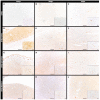
Cleaved caspase-3 immunolabeling in murine subcutaneous DH82 cell xenografts (AL). (AC) display non-infected (DH82), (DF) intratumorally CDV-infected (DH82-CDVai), (GI) intratumorally UV-inactivated CDV-injected (DH82-UV-CDVai) and (JL) intratumorally medium-injected (DH82-medium) neoplasms at (A,D,G,J) 44 days post-transplantation (dpt), (B,E,H,K) 54 dpt, and (C,F,I,L) 63 dpt. Scale bar = 100 µm and scale bar—insert = 20 µm.
Figure 4
Figure 4
Mac3 immunolabeling for assessment of tumor-associated macrophages in murine subcutaneous DH82 cell xenografts (AL). (AC) display non-infected (DH82), (DF) intratumorally canine distemper virus (CDV)-infected (DH82-CDVai), (GI) intratumorally UV-inactivated CDV-injected (DH82-UV-CDVai) and (JL) intratumorally medium-injected (DH82-medium) neoplasms at (A,D,G,J) 44 days post-transplantation (dpt), (B,E,H,K) 54 dpt and (C,F,I,L) 63 dpt. Scale bar = 100 µm and scale bar—insert = 20 µm.
Figure 5
Figure 5
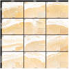
Peripheral MMP-2 immunolabeling in murine subcutaneous DH82 cell xenografts (AL). (AC) display non-infected (DH82), (DF) intratumorally canine distemper virus (CDV)-infected (DH82-CDVai), (GI) intratumorally UV-inactivated CDV-injected (DH82-UV-CDVai) and (JL) intratumorally medium-injected (DH82-medium) neoplasms at (A,D,G,J) 44 days post-transplantation (dpt), (B,E,H,K) 54 dpt and (C,F,I,L) 63 dpt. Scale bar = 100 µm and scale bar—insert = 20 µm.
Figure 6
Figure 6
Peripheral MMP-9 immunolabeling in murine subcutaneous DH82 cell xenografts (AL). (AC) display non-infected (DH82), (DF) intratumorally canine distemper virus (CDV)-infected (DH82-CDVai), (GI) intratumorally UV-inactivated CDV-injected (DH82-UV-CDVai) and (JL) intratumorally medium-injected (DH82-medium) neoplasms at (A,D,G,J) 44 days post-transplantation (dpt), (B,E,H,K) 54 dpt and (C,F,I,L) 63 dpt. Scale bar = 100 µm and scale bar—insert = 20 µm.
Figure 7
Figure 7
Peripheral MMP-14 immunolabeling in murine subcutaneous DH82 cell xenografts (AL). (AC) display non-infected (DH82), (DF) intratumorally canine distemper virus (CDV)-infected (DH82-CDVai), (GI) intratumorally UV-inactivated CDV-injected (DH82-UV-CDVai) and (JL) intratumorally medium-injected (DH82-medium) neoplasms at (A,D,G,J) 44 days post-transplantation (dpt), (B,E,H,K) 54 dpt and (C,F,I,L) 63 dpt. Scale bar = 100 µm and scale bar—insert = 20 µm.
Figure 8
Figure 8
Peripheral TIMP-1 immunolabeling in murine subcutaneous DH82 cell xenografts (AL). (AC) display non-infected (DH82), (DF) intratumorally canine distemper virus (CDV)-infected (DH82-CDVai), (GI) intratumorally UV-inactivated CDV-injected (DH82-UV-CDVai) and (JL) intratumorally medium-injected (DH82-medium) neoplasms at (A,D,G,J) 44 days post-transplantation (dpt), (B,E,H,K) 54 dpt and (C,F,I,L) 63 dpt. Scale bar = 100 µm and scale bar—insert = 20 µm.
Figure 9
Figure 9
Comparative illustration of the percentage of immunolabeled tumor tissue within the periphery of non-infected (DH82), intratumorally canine distemper virus (CDV)-infected (DH82-CDVai), intratumorally UV-inactivated CDV-injected (DH82-UV-CDVai) and intratumorally medium-injected (DH82-medium) xenografts. The graphs show the percentage of (A) matrix metalloproteinase (MMP)-2, (B) MMP-9, (C) MMP-14 and (D) their inhibitor (TIMP)-1 immunolabeling over time (days post-transplantation, dpt). All graphs represent Box and whisker plots with statistically significant differences between different groups at the same time point (** p ≤ 0.01, *** p ≤ 0.001, and **** p ≤ 0.0001) and between different time points within the same group (a–i; p < 0.05).
Figure 10
Figure 10
CD31 immunolabeling in murine subcutaneous DH82 cell xenografts for assessment of microvessel density. All CD31-expressing structures with a definable lumen were counted as vessels (arrowheads). (A) displays non-infected (DH82), (B) intratumorally canine distemper virus (CDV)-infected (DH82-CDVai), (C) intratumorally UV-inactivated CDV-injected (DH82-UV-CDVai) and (D) intratumorally medium-injected (DH82-medium) neoplasms at 63 days post-transplantation (dpt). (E) shows a graphical overview of the microvessel density within the different groups. All graphs represent Box and whisker plots with statistically significant differences between different groups at the same time point (* p < 0.05 and ** p ≤ 0.01) and between different time points within the same group (a–h; p < 0.05). Scale bar = 100 µm and scale bar—insert = 20 µm.

References

    1. O’Neill D.G., Church D.B., McGreevy P.D., Thomson P.C., Brodbelt D.C. Longevity and mortality of owned dogs in England. Vet. J. 2013;198:638–643. doi: 10.1016/j.tvjl.2013.09.020. - DOI - PubMed
    1. Ferlay J., Soerjomataram I., Dikshit R., Eser S., Mathers C., Rebelo M., Parkin D.M., Forman D., Bray F. Cancer incidence and mortality worldwide: Sources, methods and major patterns in GLOBOCAN 2012. Int. J. Cancer. 2015;136:E359–E386. doi: 10.1002/ijc.29210. - DOI - PubMed
    1. Lewis T.W., Wiles B.M., Llewellyn-Zaidi A.M., Evans K.M., O’Neill D.G. Longevity and mortality in Kennel Club registered dog breeds in the UK in 2014. Canine Genet. Epidemiol. 2018;5:10. doi: 10.1186/s40575-018-0066-8. - DOI - PMC - PubMed
    1. Bilici A. Prognostic factors related with survival in patients with pancreatic adenocarcinoma. World J. Gastroenterol. 2014;20:10802–10812. doi: 10.3748/wjg.v20.i31.10802. - DOI - PMC - PubMed
    1. Lemjabbar-Alaoui H., Hassan O.U., Yang Y.W., Buchanan P. Lung cancer: Biology and treatment options. Biochim. Biophys. Acta. 2015;1856:189–210. doi: 10.1016/j.bbcan.2015.08.002. - DOI - PMC - PubMed

MeSH terms

Substances