Review
. 2021 Mar 30;9(4):351.
doi: 10.3390/biomedicines9040351.

Potential Impact of Human Cytomegalovirus Infection on Immunity to Ovarian Tumours and Cancer Progression

Affiliations
Review

Potential Impact of Human Cytomegalovirus Infection on Immunity to Ovarian Tumours and Cancer Progression

Momodou Cox et al. Biomedicines. .

Abstract

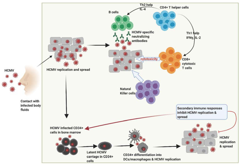
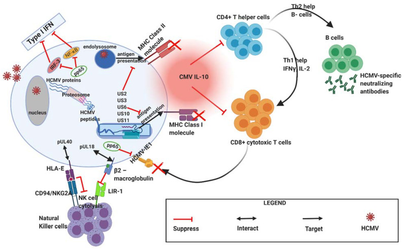
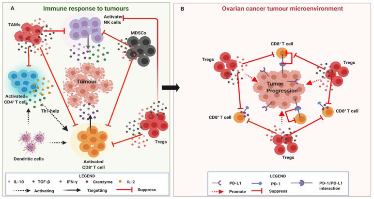
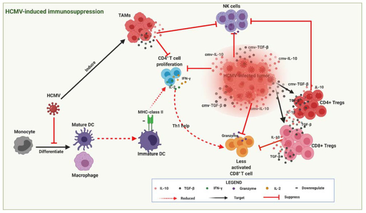
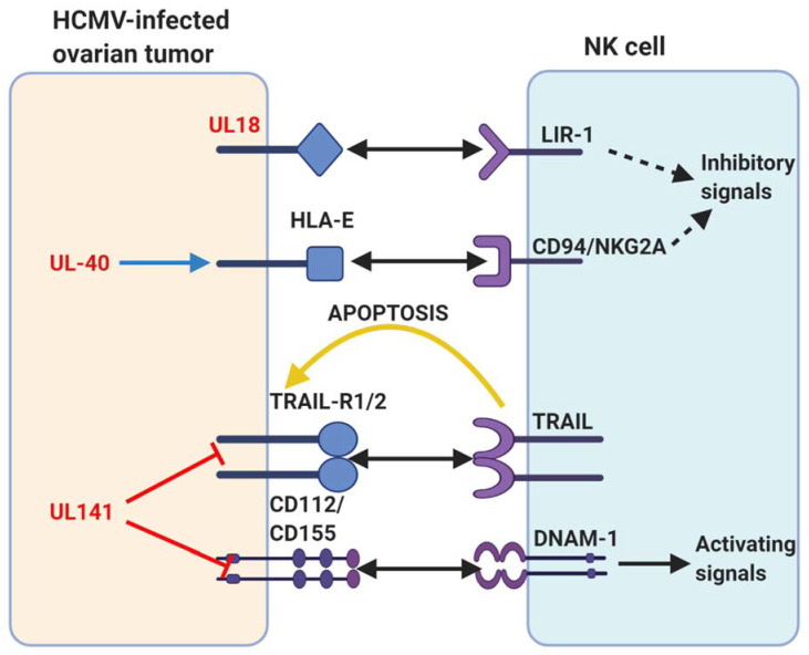
Ovarian cancer (OC) is one of the most common, and life-threatening gynaecological cancer affecting females. Almost 75% of all OC cases are diagnosed at late stages, where the 5-year survival rate is less than 30%. The aetiology of the disease is still unclear, and there are currently no screening method nor effective treatment strategies for the advanced disease. A growing body of evidence shows that human cytomegalovirus (HCMV) infecting more than 50% of the world population, may play a role in inducing carcinogenesis through its immunomodulatory activities. In healthy subjects, the primary HCMV infection is essentially asymptomatic. The virus then establishes a life-long chronic latency primarily in the hematopoietic progenitor cells in the bone marrow, with periodic reactivation from latency that is often characterized by high levels of circulating pro-inflammatory cytokines. Currently, infection-induced chronic inflammation is considered as an essential process for OC progression and metastasis. In line with this observation, few recent studies have identified high expressions of HCMV proteins on OC tissue biopsies that were associated with poor survival outcomes. Active HCMV infection in the OC tumour microenvironment may thus directly contribute to OC progression. In this review, we highlight the potential impact of HCMV infection-induced immunomodulatory effects on host immune responses to OC that may promote OC progression.

Keywords: cancer progression; human cytomegalovirus; immunosuppression; inflammation; ovarian cancer.


Conflict of interest statement

The authors declare no conflict of interest.

Figures

Figure 1
Figure 1
Following primary infection through contact with infected bodily fluids, HCMV replicates within host cells during which time robust immune responses are generated by the host that includes HCMV-specific neutralizing antibodies, natural killer cells and high frequencies of CD4+ T helper cells and CD8+ cytotoxic T cells. These responses subsequently control viral replication thereby resolving primary infection. However, HCMV has the potential to replicate and spread resulting in infection of CD34+ myeloid cells in the bone marrow and establishment of latency. Differentiation of HCMV-infected CD34+ cells into dendritic cells and macrophages contributes to new HCMV replication during which a secondary immune response induced that helps to inhibit HCMV replication spread. HCMV, human cytomegalovirus. Figure created with BioRender.com.
Figure 2
Figure 2
During primary infection, HCMV employs various mechanisms to mediate immune evasion. HCMV expresses viral genes and proteins that interfere with host interferon responses (pp65), inhibit natural killer cell recognition or activation (pUL40 and pUL18), inhibit CD4+ and CD8+ T-cell recognition by preventing MHC Class I and II antigen processing and presentation (e.g., US2, US3, US6, US10, US11). CMV IL-10 (viral IL-10 homologue) produced by infected cells further acts to suppress CD4+ and C8+ T cell responses. HCMV, human cytomegalovirus; MHC, major histocompatibility complex (MHC); US, unique short; UL, unique long. Figure created with BioRender.com.
Figure 3
Figure 3
(A)The human immune system harbours various immune cells such as natural killer (NK) cells, CD8+ T cells, CD4+ T helper cells and dendritic cells (DCs) that play an important role in controlling the developing tumour. DCs and CD4+ T cells via IL-2 and INF-γ secretion activate CD8+ cytotoxic T cells (CTLs) and NK cells which then produce toxic molecules such as granzymes that target the developing tumour. However, pro-tumour cells such as regulatory T cells (Tregs), myeloid-derived suppressor cells (MDSCs) and tumour-associated macrophages (TAMs) produce immunosuppressive cytokines (IL-10 and TGF-β) that inhibit CDTLs and NK cells effector responses. (B). The ovarian cancer tumour microenvironment is commonly characterised by high frequencies of infiltrating Tregs and a high Tregs:CD8+ T cell ratio that promotes tumour progression. Ovarian tumour also expresses PD-L1 that further inhibits CD8+ T cell effector responses upon ligation with PD-1. Figure created with BioRender.com.
Figure 4
Figure 4
HCMV-infected tumour cells secrete viral cytokines (cIL-10 and c-TGF-β) that create an immunosuppressive environment (highlighted red zone) around the developing tumour thereby promoting tumour progression. cIL-10 and c-TGF-β also induce CD4+ and CD8+ Tregs that produce similar cytokines (IL-10 and TGF-β) further augmenting the immunosuppressive state by inhibiting cytotoxic CD8+ T lymphocyte (CTL) and natural killer (NK) cell effector responses. HCMV also interferes with the differentiation of monocytes into mature dendritic cells (DCs) that leads to the establishment of immature DCs with reduced MHC-class II antigen presentation and subsequently less CD4+ T cell proliferation and reduced Th1 help needed for enhanced CTL activation. Figure created with BioRender.com.
Figure 5
Figure 5
A summary of human cytomegalovirus (HCMV) modulation of natural killer (NK) cell cytotoxicity discussed in this review. Human proteins or receptors are labelled in black; HCMV proteins are labelled in red. The arrows represent actions. Two-pointed solid black arrow = interaction or ligation; one-pointed solid black arrow = intracellular NK cell activation signal; dotted black arrow = intracellular NK cell inhibition signal; yellow arrow = extracellular signal to target cell; red line = inhibits surface expression; blue arrow = increases surface expression. Figure created with BioRender.com.

References

    1. Sankaranarayanan R., Ferlay J. Worldwide burden of gynaecological cancer: The size of the problem. Best Pr. Res. Clin. Obstet. Gynaecol. 2006;20:207–225. doi: 10.1016/j.bpobgyn.2005.10.007. - DOI - PubMed
    1. Reid B.M., Permuth J.B., Sellers T.A. Epidemiology of ovarian cancer: A review. Cancer Biol. Med. 2017;14:9–32. doi: 10.20892/j.issn.2095-3941.2016.0084. - DOI - PMC - PubMed
    1. Xie X., Yang M., Ding Y., Chen J. Microbial infection, inflammation and epithelial ovarian cancer. Oncol. Lett. 2017;14:1911–1919. doi: 10.3892/ol.2017.6388. - DOI - PMC - PubMed
    1. Kampan N.C., Madondo M.T., McNally O.M., A Quinn M., Plebanski M. Paclitaxel and Its Evolving Role in the Management of Ovarian Cancer. BioMed. Res. Int. 2015;2015:1–21. doi: 10.1155/2015/413076. - DOI - PMC - PubMed
    1. Aust S., Seebacher-Shariat V. Screening for ovarian cancer: Is there still hope? MEMO Mag. Eur. Med. Oncol. 2020;13:189–192. doi: 10.1007/s12254-019-00563-2. - DOI

LinkOut - more resources