. 2021 Mar 30;26(7):1937.
doi: 10.3390/molecules26071937.

The Ameliorative Effects of Fucoidan in Thioacetaide-Induced Liver Injury in Mice

Affiliations

The Ameliorative Effects of Fucoidan in Thioacetaide-Induced Liver Injury in Mice

Ming-Yang Tsai et al. Molecules. .

Abstract

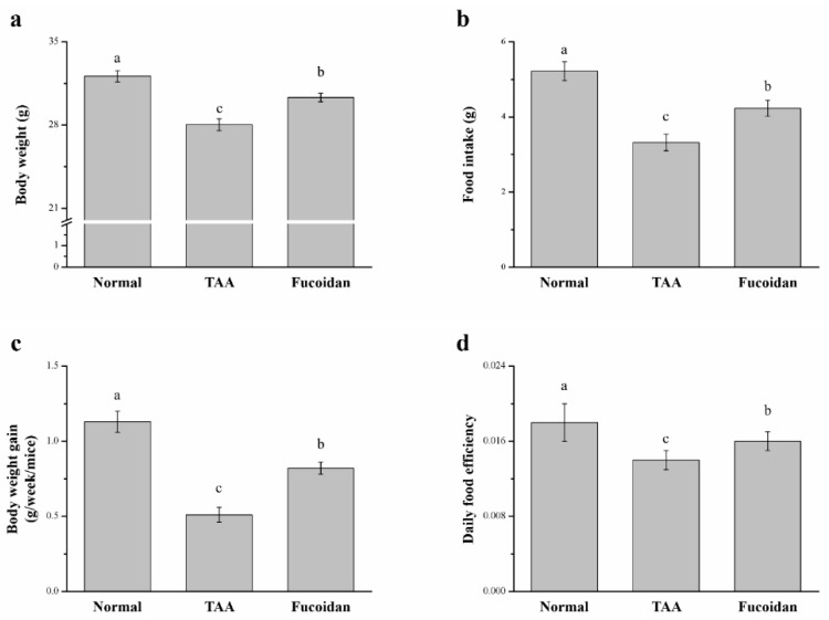
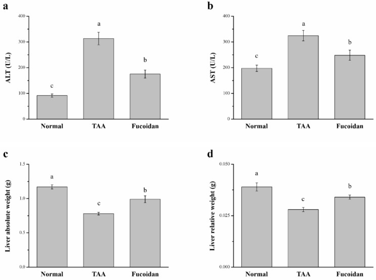
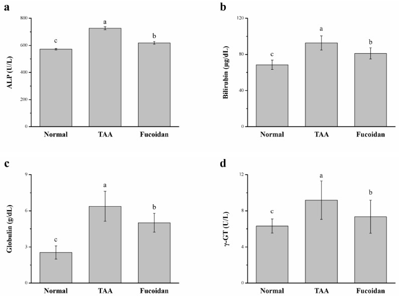
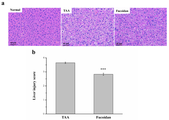
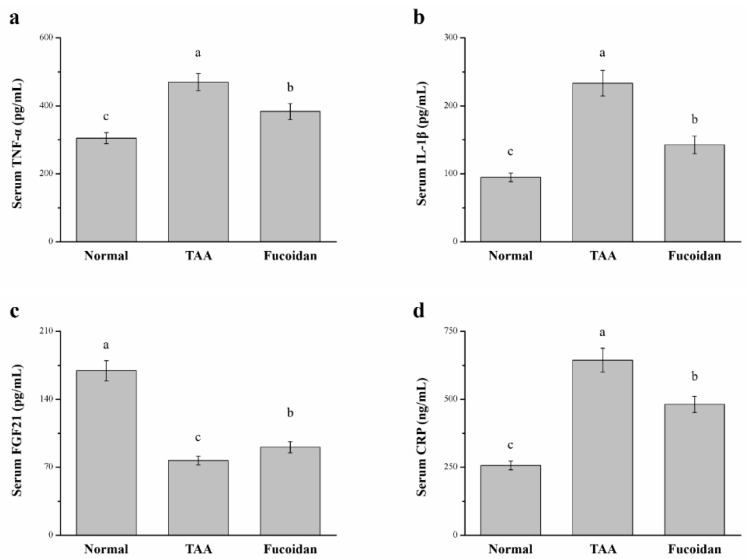
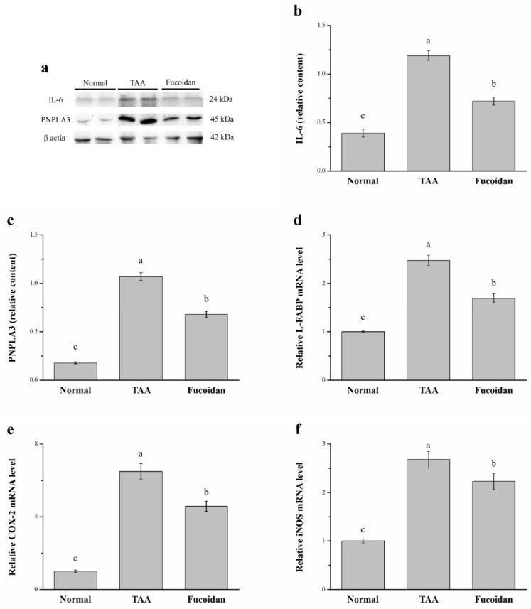
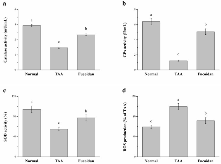
Liver disorders have been recognized as one major health concern. Fucoidan, a sulfated polysaccharide extracted from the brown seaweed Fucus serratus, has previously been reported as an anti-inflammatory and antioxidant. However, the discovery and validation of its hepatoprotective properties and elucidation of its mechanisms of action are still unknown. The objective of the current study was to investigate the effect and possible modes of action of a treatment of fucoidan against thioacetamide (TAA)-induced liver injury in male C57BL/6 mice by serum biochemical and histological analyses. The mouse model for liver damage was developed by the administration of TAA thrice a week for six weeks. The mice with TAA-induced liver injury were orally administered fucoidan once a day for 42 days. The treated mice showed significantly higher body weights; food intakes; hepatic antioxidative enzymes (catalase, glutathione peroxidase (GPx), and superoxide dismutase (SOD)); and a lower serum alanine aminotransferase (ALT), aspartate aminotransferase (AST), tumor necrosis factor-α (TNF-α), interleukin-1β (IL-1β), and C-reactive protein (CRP) levels. Additionally, a reduced hepatic IL-6 level and a decreased expression of inflammatory-related genes, such as cyclooxygenase-2 (COX-2), and inducible nitric oxide synthase (iNOS) mRNA was observed. These results demonstrated that fucoidan had a hepatoprotective effect on liver injury through the suppression of the inflammatory responses and acting as an antioxidant. In addition, here, we validated the use of fucoidan against liver disorders with supporting molecular data.

Keywords: fucoidan; inflammation; liver; mice; thioacetamide.


Conflict of interest statement

The authors declare no conflict of interest.

Figures

Figure 1
Figure 1
Changes of the (a) body weight, (b) food intake, (c) weekly body weight gain, and (d) daily food efficiency in normal mice, mice with thioacetamide (TAA)-induced liver injury, and fucoidan-treated mice with TAA-induced liver injury within a time period of 42 days. All data are presented as the means ± standard error (SEMs) (n = 10). a–c Data with different letters in the columns are significantly different with one-way analysis of variance (ANOVA), and the means of different groups were compared by Duncan’s test at p < 0.05.
Figure 2
Figure 2
Changes in the (a) serum alanine aminotransferase (ALT) levels, (b) serum aspartate aminotransferase (AST) levels, (c) absolute weights, and (d) body weight-normalized weights (%) of the livers in normal mice, mice with TAA-induced liver injury, and fucoidan-treated mice with TAA-induced liver injury over a period of 42 days. All data are presented as the means ± SEMs (n = 10). a–c Data with different letters in the columns are significantly different with one-way ANOVA, and the means of different groups were compared by Duncan’s test at p < 0.05.
Figure 3
Figure 3
Changes of the serum (a) alkaline phosphatase (ALP), (b) bilirubin, (c) globulin, and (d) γ-glutamyl transferase (γ-GT) levels in normal mice, in mice with TAA-induced liver injury, and fucoidan-treated mice with TAA-induced liver injury over a period of 42 days. All data are presented as the means ± SEMs (n = 10). a–c Data with different letters in the columns are significantly different with one-way ANOVA, and the means of the different groups were compared by Duncan’s test at p < 0.05.
Figure 4
Figure 4
Changes of the (a) liver histology by hematoxylin and eosin (H&E) staining (magnification 200×), and (b) the scoring index of liver injuries in normal mice, mice with TAA-induced liver injury, and fucoidan-treated mice with TAA-induced liver injury over a period of 42 days. All data are presented as the means ± SEMs (n = 10). Statistical significance at *** p < 0.001.
Figure 5
Figure 5
Changes of the serum (a) tumor necrosis factor-α (TNF-α), (b) interleukin-1β (IL-1β), (c) fibroblast growth factor-21 (FGF21), and (d) C-reactive protein (CRP) levels in normal mice, mice with TAA-induced liver injury, and fucoidan-treated mice with TAA-induced liver injury after 42 days. All data are presented as the means ± SEMs (n = 10). a–c Data with different letters in the columns are significantly different with one-way ANOVA, and the means of different groups were compared by Duncan’s test at p < 0.05.
Figure 6
Figure 6
(a) Representative Western blot of the liver extracts for interleukin-6 (IL-6) and patatin-like phospholipid domain containing protein 3 (PNPLA3) expression, (b) IL-6, and (c) PNPLA3 expression, as well as changes in the (d) liver fatty acid-binding protein (L-FABP), (e) cyclooxygenase-2 (COX-2), and (f) inducible nitric oxide synthase (iNOS) mRNA levels in the livers of normal mice, TAA-induced liver injury mice, and fucoidan-treated TAA-induced liver injury mice over a time period of 42 days. All data are presented as the means ± SEMs (n = 10). a–c Data with different letters in the columns are significantly different with one-way ANOVA, and the means of different groups were compared by Duncan’s test at p < 0.05.
Figure 7
Figure 7
Changes of the hepatic (a) catalase activity, (b) glutathione peroxidase (GPx) activity, (c) superoxide dismutase (SOD) activity, and (d) reactive oxygen species (ROS) levels in normal mice, mice with TAA-induced liver injury, and fucoidan-treated mice with TAA-induced liver injury over a period of 42 days. All data are presented as the means ± SEMs (n = 10). a–c Data with different letters in the columns are significantly different with one-way ANOVA, and the means of different groups were compared by Duncan’s test at p < 0.05.

References

    1. Treyer A., Müsch A. Hepatocyte polarity. Compr. Physiol. 2013;3:243–287. - PMC - PubMed
    1. Blouin A., Bolender R.P., Weibel E.R. Distribution of organelles and memebranes between hepatocytes and nonhepatocytes in the rat liver parenchyma. A stereological study. Cell Biol. 1977;72:441–455. doi: 10.1083/jcb.72.2.441. - DOI - PMC - PubMed
    1. De Mingo Pulido Á., de Gregorio E., Chandra S., Colell A., Morales A., Kronenberg M., Marí M. Differential role of cathepsins S and B in hepatic APC-mediated NKT cell activation and cytokine secretion. Front. Immunol. 2018;9:391. doi: 10.3389/fimmu.2018.00391. - DOI - PMC - PubMed
    1. Hirsova P., Gores G.J. Death receptor-mediated cell death and proinflammatory signaling in nonalcoholic steatohepatitis. Cell. Mol. Gastroenterol. Hepatol. 2015;1:17–27. doi: 10.1016/j.jcmgh.2014.11.005. - DOI - PMC - PubMed
    1. Yang L., Seki E. Toll-like receptors in liver fibrosis: Cellular crosstalk and mechanisms. Front. Physiol. 2012;3:138. doi: 10.3389/fphys.2012.00138. - DOI - PMC - PubMed

MeSH terms