Review
. 2021 Mar 30;22(7):3591.
doi: 10.3390/ijms22073591.

The Post-Translational Regulation of Epithelial-Mesenchymal Transition-Inducing Transcription Factors in Cancer Metastasis

Affiliations
Review

The Post-Translational Regulation of Epithelial-Mesenchymal Transition-Inducing Transcription Factors in Cancer Metastasis

Eunjeong Kang et al. Int J Mol Sci. .

Abstract

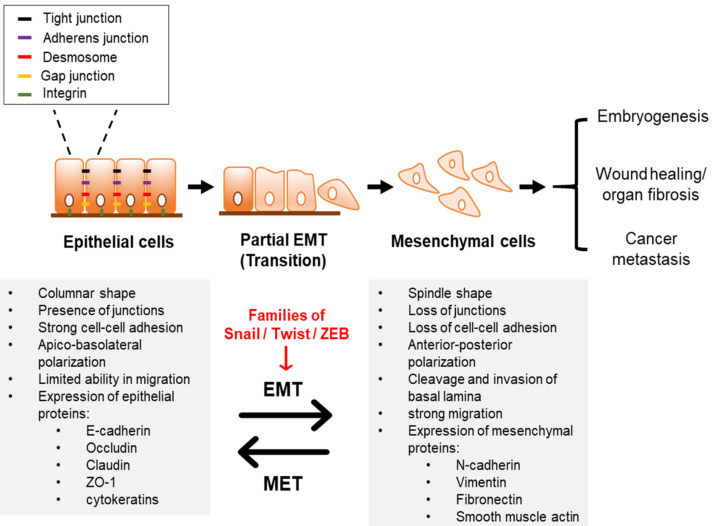
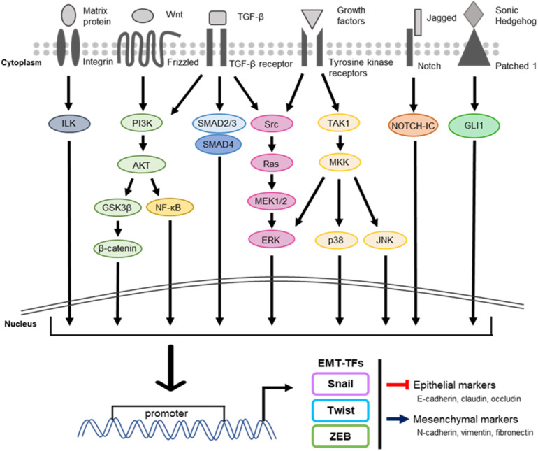
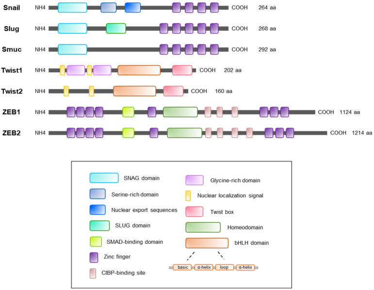
Epithelial-mesenchymal transition (EMT) is generally observed in normal embryogenesis and wound healing. However, this process can occur in cancer cells and lead to metastasis. The contribution of EMT in both development and pathology has been studied widely. This transition requires the up- and down-regulation of specific proteins, both of which are regulated by EMT-inducing transcription factors (EMT-TFs), mainly represented by the families of Snail, Twist, and ZEB proteins. This review highlights the roles of key EMT-TFs and their post-translational regulation in cancer metastasis.

Keywords: Snail; Twist; ZEB; epithelial–mesenchymal transition; metastasis; transcription factor.


Conflict of interest statement

The authors declare no conflict of interest.

Figures

Figure 1
Figure 1
Characteristics of EMT. Epithelial cells are usually attached to the basement membrane. These cells maintain cell–cell connection such as adherens, tight and gap junctions, and desmosomes. EMT is primarily involved in normal embryogenesis and is associated with adult tissue regeneration, wound healing, and fibrosis. As EMT of cancer cells is transient, the mesenchymal state of cells reverts to the epithelial phenotype, which is called as MET. The process of EMT causes epithelial markers (e.g., E-cadherin, claudin, ZO-1) to be gradually lost, while mesenchymal markers (e.g., N-cadherin, vimentin, and fibronectin) to be increased, which causes changes in physiology of cells. Hence, cells acquire high motility and invasive properties. This process is regulated by EMT-TFs such as families of Snail, Twist, and ZEB. EMT, epithelial–mesenchymal transition; EMT-TFs, EMT-inducing transcription factors; MET, mesenchymal–epithelial transition; ZO-1, Zonula occludens-1.
Figure 2
Figure 2
The common signaling pathways regulating EMT. The progression of EMT is controlled by several extracellular and intracellular signaling pathways. Their coordinated interactions bind to DNA promoter regions of EMT-TFs, leading to promotion of transcriptional activity of EMT-TFs. The expressions of EMT-TFs play a key role in regulating the expression of their target genes related to EMT and cancer metastasis. (Left to right: matrix protein/ILK, WNT/PI3K/β-catenin, TGF-β/PI3K/NF-κB, TGF-β/SMAD complex, growth factors or TGF-β/RAS/ERK, growth factors/TAK1/MAPK, Jagged/NOTCH-ICD, and Sonic Hedgehog/GLI1) ILK, integrin-linked kinase; TGF-β, transforming growth factor-beta; PI3K, phosphoinositide 3-kinase; NF-κB, nuclear factor-kappa B; ERK, extracellular signal-regulated kinase; JNK, c-Jun N-terminal kinases; MAPK, mitogen-activated protein kinase; TAK1, transforming growth factor β-activated kinase 1; MEK, MAPK/ERK kinase; MKK, mitogen-activated protein kinase kinase.
Figure 3
Figure 3
A schematic representation of structures of EMT-TFs. The Snail family (Snail, Slug, and Smuc) commonly includes the SNAG domain in the N-terminal and the zinc finger domain in the C-terminal. Snail contains the nuclear export sequences and serine-rich domain that control the stability of the Snail. The SNAG domain is however only present in Slug. The latest recognized member in Snail family, Smuc does not have both serine-rich domain and SNAG domain. The Twist family (Twist1 and Twist2) mainly consists of a bHLH domain and a Twist box in C-terminal for its transcriptional activity. However, glycine rich domains are only present in Twist1. The bHLH domain consists of basic amino acids followed by two alpha helices, which is separated by a loop of different length. The ZEB family (ZEB1 and ZEB2) has the largest protein size compared to other EMT-TFs. This ZEB family contains various regulatory domains, which include clusters of zinc fingers in N-terminal and C-terminal, homeodomain, SMAD-binding domain, and CtBP-binding site. CtBP, C-terminal binding protein.

References

    1. Li L., Li W. Epithelial-mesenchymal transition in human cancer: Comprehensive reprogramming of metabolism, epigenetics, and differentiation. Pharmacol. Ther. 2015;150:33–46. doi: 10.1016/j.pharmthera.2015.01.004. - DOI - PubMed
    1. Škovierová H., Okajčeková T., Strnádel J., Vidomanová E., Halašová E. Molecular regulation of epithelial-to-mesenchymal transition in tumorigenesis (Review) Int. J. Mol. Med. 2018;41:1187–1200. doi: 10.3892/ijmm.2017.3320. - DOI - PMC - PubMed
    1. Garg M. Epithelial-mesenchymal transition-activating transcription factors-multifunctional regulators in cancer. World J. Stem Cells. 2013;5:188. doi: 10.4252/wjsc.v5.i4.188. - DOI - PMC - PubMed
    1. Stone R.C., Pastar I., Ojeh N., Chen V., Liu S., Garzon K.I., Tomic-Canic M. Epithelial-mesenchymal transition in tissue repair and fibrosis. Cell Tissue Res. 2016;365:495–506. doi: 10.1007/s00441-016-2464-0. - DOI - PMC - PubMed
    1. Navas T., Kinders R.J.J., Lawrence S.M.M., Ferry-Galow K.V.V., Borgel S., Hollingshead M.G.G., Srivastava A.K.K., Alcoser S.Y.Y., Makhlouf H.R.R., Chuaqui R., et al. Clinical evolution of epithelial–mesenchymal transition in human carcinomas. Cancer Res. 2020;80:304–318. doi: 10.1158/0008-5472.CAN-18-3539. - DOI - PMC - PubMed

MeSH terms

Substances

LinkOut - more resources