

Implication of the NLRP3 Inflammasome in Bovine Age-Related Sarcopenia
- PMID: 33808510
- PMCID: PMC8036417
- DOI: 10.3390/ijms22073609
Implication of the NLRP3 Inflammasome in Bovine Age-Related Sarcopenia
Abstract
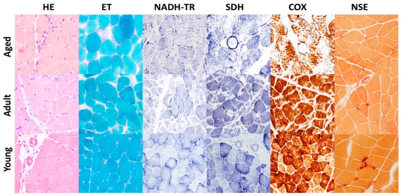
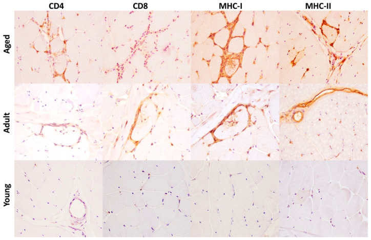
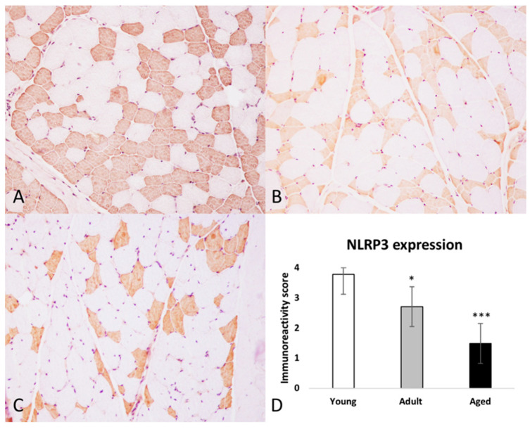
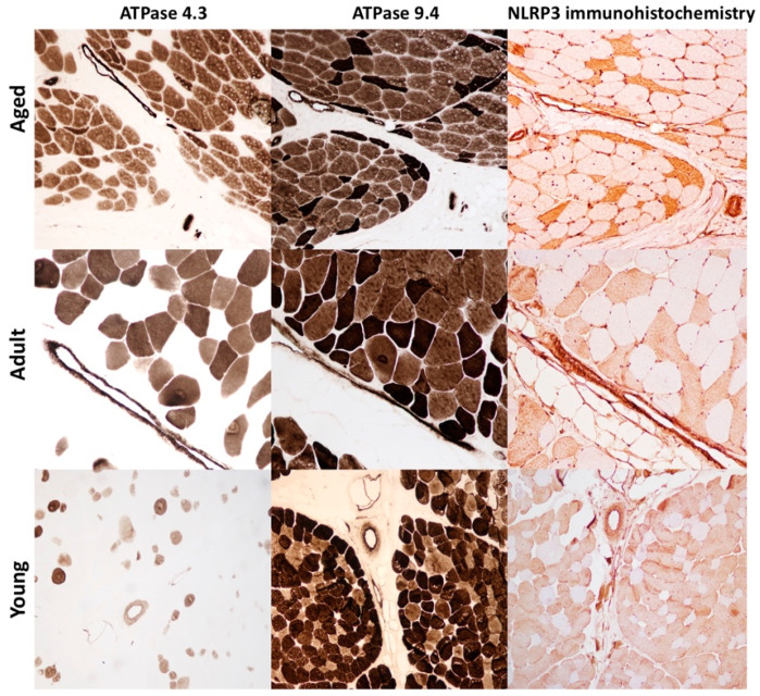
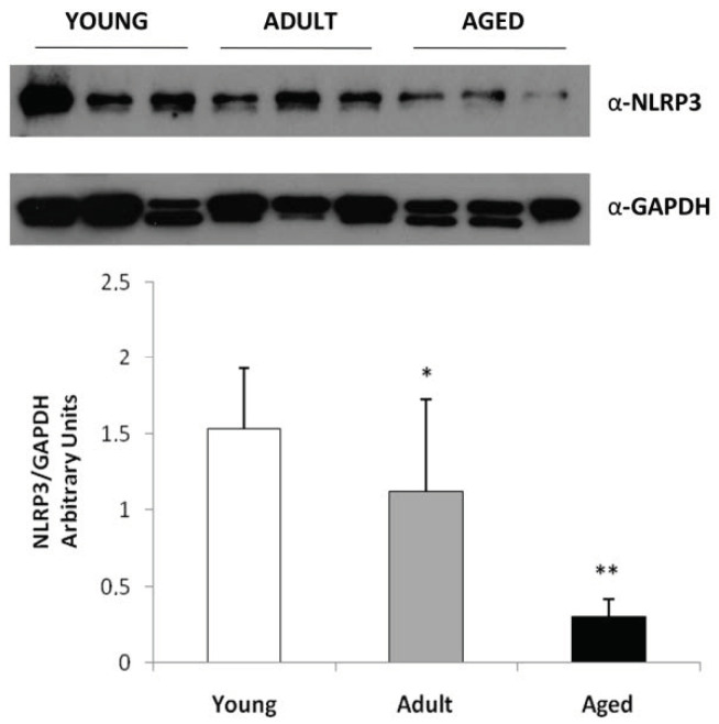
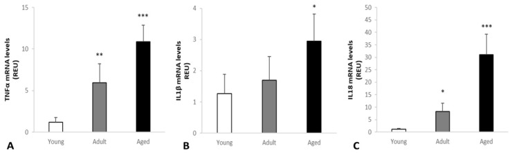
Sarcopenia is defined as the age-related loss of skeletal muscle mass, quality, and strength. The pathophysiological mechanisms underlying sarcopenia are still not completely understood. The aim of this work was to evaluate, for the first time, the expression of NLRP3 inflammasome in bovine skeletal muscle in order to investigate the hypothesis that inflammasome activation may trigger and sustain a pro-inflammatory environment leading to sarcopenia. Samples of skeletal muscle were collected from 60 cattle belonging to three age-based groups. Morphologic, immunohistochemical and molecular analysis were performed to assess the presence of age-related pathologic changes and chronic inflammation, the expression of NLRP3 inflammasome and to determine the levels of interleukin-1β, interleukin-18 and tumor necrosis factor alpha in muscle tissue. Our results revealed the presence of morphologic sarcopenia hallmark, chronic lymphocytic inflammation and a type II fibers-selective NLRP3 expression associated to a significant decreased number of immunolabeled-fibers in aged animals. Moreover, we found a statistically significant age-related increase of pro-inflammatory cytokines such as interleukin-1β and interleukin-18 suggesting the activation of NLRP3 inflammasome. Taken together, our data suggest that NLRP3 inflammasome components may be normally expressed in skeletal muscle, but its priming and activation during aging may contribute to enhance a pro-inflammatory environment altering normal muscular anabolism and metabolism.
Keywords: NLRP3 inflammasome; immunosenescence; inflammaging; sarcopenia.
Conflict of interest statement
The authors declare no conflict of interest.
Figures
Similar articles
-
NLRP3 Contributes to Sarcopenia Associated to Dependency Recapitulating Inflammatory-Associated Muscle Degeneration.Int J Mol Sci. 2024 Jan 24;25(3):1439. doi: 10.3390/ijms25031439. Int J Mol Sci. 2024. PMID: 38338718 Free PMC article.
-
The NLRP3 inflammasome contributes to sarcopenia and lower muscle glycolytic potential in old mice.Am J Physiol Endocrinol Metab. 2017 Aug 1;313(2):E222-E232. doi: 10.1152/ajpendo.00060.2017. Epub 2017 May 23. Am J Physiol Endocrinol Metab. 2017. PMID: 28536183 Free PMC article.
-
Hydrogen-Rich Saline Attenuated Subarachnoid Hemorrhage-Induced Early Brain Injury in Rats by Suppressing Inflammatory Response: Possible Involvement of NF-κB Pathway and NLRP3 Inflammasome.Mol Neurobiol. 2016 Jul;53(5):3462-3476. doi: 10.1007/s12035-015-9242-y. Epub 2015 Jun 20. Mol Neurobiol. 2016. PMID: 26091790
-
New insights into the function of the NLRP3 inflammasome in sarcopenia: mechanism and therapeutic strategies.Metabolism. 2024 Sep;158:155972. doi: 10.1016/j.metabol.2024.155972. Epub 2024 Jul 6. Metabolism. 2024. PMID: 38972476 Review.
-
[Advances in mechanisms for NLRP3 inflammasomes regulation].Yao Xue Xue Bao. 2016 Oct;51(10):1505-12. Yao Xue Xue Bao. 2016. PMID: 29924571 Review. Chinese.
Cited by
-
Pyroptosis and Sarcopenia: Frontier Perspective of Disease Mechanism.Cells. 2022 Mar 23;11(7):1078. doi: 10.3390/cells11071078. Cells. 2022. PMID: 35406642 Free PMC article. Review.
-
Evaluation of Muscle Proteins for Estimating the Post-Mortem Interval in Veterinary Forensic Pathology.Animals (Basel). 2023 Feb 6;13(4):563. doi: 10.3390/ani13040563. Animals (Basel). 2023. PMID: 36830350 Free PMC article.
-
Association between serum uric acid, hyperuricemia and low muscle mass in middle-aged and elderly adults: A national health and nutrition examination study.PLoS One. 2025 Jan 7;20(1):e0312235. doi: 10.1371/journal.pone.0312235. eCollection 2025. PLoS One. 2025. PMID: 39775063 Free PMC article.
-
Pathological alterations and COHb evaluations as tools for investigating fire-related deaths in veterinary forensic pathology.Front Vet Sci. 2024 May 21;11:1396540. doi: 10.3389/fvets.2024.1396540. eCollection 2024. Front Vet Sci. 2024. PMID: 38835893 Free PMC article.
References
MeSH terms
Substances
LinkOut - more resources
Full Text Sources
Other Literature Sources
