

Artificial Intelligence (AI)-Empowered Echocardiography Interpretation: A State-of-the-Art Review
- PMID: 33808513
- PMCID: PMC8037652
- DOI: 10.3390/jcm10071391
Artificial Intelligence (AI)-Empowered Echocardiography Interpretation: A State-of-the-Art Review
Abstract
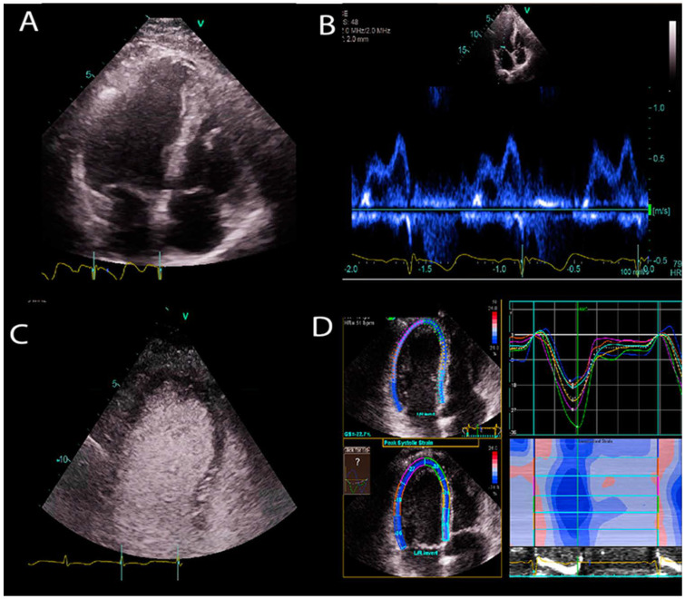
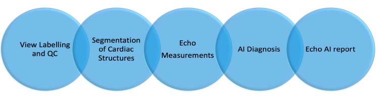
Echocardiography (Echo), a widely available, noninvasive, and portable bedside imaging tool, is the most frequently used imaging modality in assessing cardiac anatomy and function in clinical practice. On the other hand, its operator dependability introduces variability in image acquisition, measurements, and interpretation. To reduce these variabilities, there is an increasing demand for an operator- and interpreter-independent Echo system empowered with artificial intelligence (AI), which has been incorporated into diverse areas of clinical medicine. Recent advances in AI applications in computer vision have enabled us to identify conceptual and complex imaging features with the self-learning ability of AI models and efficient parallel computing power. This has resulted in vast opportunities such as providing AI models that are robust to variations with generalizability for instantaneous image quality control, aiding in the acquisition of optimal images and diagnosis of complex diseases, and improving the clinical workflow of cardiac ultrasound. In this review, we provide a state-of-the art overview of AI-empowered Echo applications in cardiology and future trends for AI-powered Echo technology that standardize measurements, aid physicians in diagnosing cardiac diseases, optimize Echo workflow in clinics, and ultimately, reduce healthcare costs.
Keywords: artificial intelligence; cardiac ultrasound; echocardiography; portable ultrasound.
Conflict of interest statement
The authors declare no conflict of interest.
Figures

References
-
- Vincent P., Larochelle H., Lajoie I., Bengio Y., Manzagol P.-A. Stacked Denoising Autoencoders: Learning Useful Representations in a Deep Network with a Local Denoising Criterion. J. Mach. Learn. Res. 2010;11:3371–3408.
Publication types
LinkOut - more resources
Full Text Sources
Other Literature Sources
