. 2021 Mar 19;9(3):278.
doi: 10.3390/vaccines9030278.

K48-Linked Ubiquitination Contributes to Nicotine-Augmented Bone Marrow-Derived Dendritic-Cell-Mediated Adaptive Immunity

Affiliations

K48-Linked Ubiquitination Contributes to Nicotine-Augmented Bone Marrow-Derived Dendritic-Cell-Mediated Adaptive Immunity

Chun Fang Hu et al. Vaccines (Basel). .

Abstract

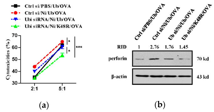
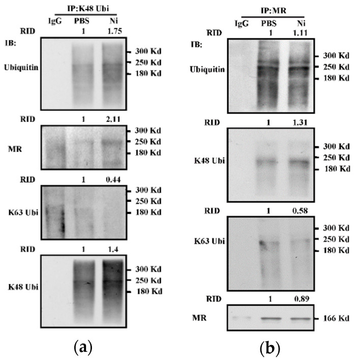
K48-linked ubiquitination determining antigen degradation and the endosomal recruitments of p97 and Sec61 plays vital roles in dendritic cell (DC) cross-presentation. Our previous studies revealed that nicotine treatment increases bone marrow-derived dendritic cell (BM-DC) cross-presentation and promotes BM-DC-based cytotoxic T lymphocyte (CTL) priming. But the effect of nicotine on K48-linked ubiquitination and the mechanism of nicotine-increased BM-DC cross-presentation are still uncertain. In this study, we first demonstrated that ex vivo nicotine administration obviously increased K48-linked ubiquitination in BM-DC. Then, we found that K48-linked ubiquitination was essential for nicotine-augmented cross-presentation, BM-DC-based CTL priming, and thereby the superior cytolytic capacity of DC-activated CTL. Importantly, K48-linked ubiquitination was verified to be necessary for nicotine-augmented endosomal recruitments of p97 and Sec61. Importantly, mannose receptor (MR), which is an important antigenic receptor for cross-presentation, was exactly catalyzed with K48-linked ubiquitination by the treatment with nicotine. Thus, these data suggested that K48-linked ubiquitination contributes to the superior adaptive immunity of nicotine-administrated BM-DC. Regulating K48-linked ubiquitination might have therapeutic potential for DC-mediated immune therapy.

Keywords: K48 ubiquitination; adaptive immunity; bone marrow precursor cells; dendritic cells; nicotine; ubiquitin.


Conflict of interest statement

The authors declare no conflict of interest.

Figures

Figure 1
Figure 1
The treatment with nicotine increases K48-linked ubiquitination in bone marrow-derived dendritic cells. (ad) Murine BM-DC was incubated with nicotine (10−7 mol/L) for 12~16 h in the presence of MG132 (50 × 10−6 mol/L). Whole cellular protein was extracted and used as input control (a). The levels of ubiquitination (b), K48-linked ubiquitination (c), and K63-linked ubiquitination (d) were determined by IP with indicated antibodies. Protein level was quantified by ImageJ software and presented as related integrated density (RID). One representative from three independent experiments is shown. Ni: nicotine; Ubi: ubiquitination; K48 Ubi: K48-linked ubiquitination; K63 Ubi: K63-linked ubiquitination; IP: immunoprecipitation; IB: immunoblotting.
Figure 2
Figure 2
K48-linked ubiquitination contributes to nicotine-increased cross-presentation in bone-marrow-derived dendritic cells. (ac) Murine ubiquitin-deficient and control BM-DC were stimulated with PBS (a) or nicotine (10−7 mol/L) (b) for 12~16 h. Then, the cells were replenished with K48R-Ub, K63R-Ub, or WT-Ub (30 × 10−6 mol/L or indicated concentration) prior to further incubation with ovalbumin (50 μg/mL). Cross-presented OVA was assessed by Western blot analyses (a) or confocal microscope (b,c). Cross-presented OVA was stained with 25-D1.16 (red) and endosome was stained with EEA1 antibody (green) respectively; nuclei were counterstained with DAPI (blue). The co-localized spots of 25-D1.16 with EEA1 were counted and analyzed (c). For Western blot analyses, β-actin was used as an internal control. Protein level was quantified by ImageJ software and presented as related integrated density (RID). Data are presented as the mean ± SEM, *** p < 0.001, one-way ANOVA with Newman-Keulspost test. One representative from three independent experiments is shown. Ni: nicotine; Ub: ubiquitin; si: siRNA; EEA1: early endosome marker; 25D1.16: antibody specific to SIINFEKL-H2Kb complex; OVA: ovalbumin.
Figure 3
Figure 3
K48-linked ubiquitination contributes to nicotine-augmented dendritic cell-based CTL priming. (a,b) Murine ubiquitin deficient and control BM-DC were stimulated with nicotine (10−7 mol/L) for 12~16 h. Then, the cells were replenished with K48R-Ub or WT-Ub (30 × 10−6 mol/L) and further incubated with ovalbumin (50 μg/mL) for 5~6 h. After that, 1 × 10⁵ BM-DC was intraperitoneally transferred into C57BL/6 recipients. 5~7 d after adoptive transfer, splenocytes and lymph nodes of the recipients were prepared. Antigen-specific CTL in the lymph nodes was determined by IFN-γ Elispot assay (a). IFN-γ expression in splenocytes was determined by Western blot analyses (b). For Western blot analyses, β-actin was used as an internal control. Protein level was quantified by ImageJ software and presented as related integrated density (RID). One representative from three independent experiments is shown. Data were presented as the mean ± SEM of replicates from one experiment. *** p < 0.001, one-way ANOVA with Newman-Keulspost test. Ni: nicotine; Ub: ubiquitin; si: siRNA; OVA: ovalbumin.
Figure 4
Figure 4
K48 ubiquitination contributes to nicotine-increased cytolytic activities of dendritic cell-activated T cells. (a,b) Murine ubiquitin deficient and control BM-DC were stimulated with nicotine (10−7 mol/L) for 12~16 h. Then, the cells were replenished with K48R-Ub or WT-Ub (30 × 10−6 mol/L) and further incubated with ovalbumin (50 μg/mL) for 5~6 h. Then, 1 × 10⁵ BM-DC was intraperitoneally transferred into C57BL/6 recipients. 5~7 d after adoptive transfer, splenocytes of the recipients were prepared and the cytolytic activities were assessed by incubation of effector cells with CFSE-labeled E.G7-OVA target cells. CFSE value was analyzed by flow cytometry, and the cytolytic activities were calculated (a). Perforin expression in the splenocytes of the recipients was determined by Western blot analyses (b). For Western blot analyses, β-actin was used as an internal control. Protein level was quantified by ImageJ software and presented as related integrated density (RID). One representative from three independent experiments is shown. Data were presented as the mean ± SEM of replicates from one experiment. * p < 0.05, *** p < 0.001, two-way ANOVA with Newman-Keulspost test. Ni: nicotine; Ub: ubiquitin; si: siRNA; OVA: ovalbumin.
Figure 5
Figure 5
K48-linked ubiquitination is essential for nicotine-augmented endosomal relocation of p97 in bone marrow-derived dendritic cells. (a,b) Murine ubiquitin-deficient and control BM-DC were stimulated with nicotine (10−7 mol/L) for 12~16 h. Then, the cells were replenished with K48R-Ub or WT-Ub (30 × 10−6 mol/L) and further incubated with ovalbumin (50 μg/mL) for 5~6 h. The endosomal relocation of p97 (a) from the endoplasmic reticulum (b) was determined by confocal microscope with EEA1, calnexin, and p97 antibody staining. EEA1 (a), calnexin (b) were stained green; p97 was stained red; nuclei were counterstained with DAPI (blue). The co-localized spots of p97 with EEA1 (c) and p97 with calnexin (d) were counted and analyzed. Data are presented as the mean ± SEM, ** p < 0.01, *** p < 0.001, one-way ANOVA with Newman-Keulspost test. One representative from three independent experiments is shown. EEA1: early endosome marker; Calnexin: endoplasmic reticulum marker; Ni: nicotine; Ub: ubiquitin; si: siRNA; OVA: ovalbumin.
Figure 5
Figure 5
K48-linked ubiquitination is essential for nicotine-augmented endosomal relocation of p97 in bone marrow-derived dendritic cells. (a,b) Murine ubiquitin-deficient and control BM-DC were stimulated with nicotine (10−7 mol/L) for 12~16 h. Then, the cells were replenished with K48R-Ub or WT-Ub (30 × 10−6 mol/L) and further incubated with ovalbumin (50 μg/mL) for 5~6 h. The endosomal relocation of p97 (a) from the endoplasmic reticulum (b) was determined by confocal microscope with EEA1, calnexin, and p97 antibody staining. EEA1 (a), calnexin (b) were stained green; p97 was stained red; nuclei were counterstained with DAPI (blue). The co-localized spots of p97 with EEA1 (c) and p97 with calnexin (d) were counted and analyzed. Data are presented as the mean ± SEM, ** p < 0.01, *** p < 0.001, one-way ANOVA with Newman-Keulspost test. One representative from three independent experiments is shown. EEA1: early endosome marker; Calnexin: endoplasmic reticulum marker; Ni: nicotine; Ub: ubiquitin; si: siRNA; OVA: ovalbumin.
Figure 6
Figure 6
K48-linked ubiquitination contributes to nicotine-increased endosomal relocation of Sec61α in dendritic cells. (a,b) Murine ubiquitin-deficient and control BM-DC were stimulated with nicotine (10−7 mol/L) for 12~16 h. Then, the cells were replenished with K48R-Ub or WT-Ub (30 × 10−6 mol/L) and further incubated with ovalbumin (50 μg/mL) for 5~6 h. The endosomal relocation of Sec61α (a) from the endoplasmic reticulum (b) was determined by confocal microscope with EEA1, calnexin, and Sec61α antibody staining. EEA1 (a), calnexin (b) were stained green; Sec61α was stained red; nuclei were counterstained with DAPI (blue). The co-localized spots of Sec61α with EEA1 (c), Sec61α with calnexin (d) were counted and analyzed. Data are presented as the mean ± SEM, * p < 0.05, *** p < 0.001, one-way ANOVA with Newman-Keulspost test. One representative from three independent experiments is shown. EEA1: early endosome marker; Calnexin: endoplasmic reticulum marker; Ni: nicotine; Ub: ubiquitin; si: siRNA; OVA: ovalbumin.
Figure 6
Figure 6
K48-linked ubiquitination contributes to nicotine-increased endosomal relocation of Sec61α in dendritic cells. (a,b) Murine ubiquitin-deficient and control BM-DC were stimulated with nicotine (10−7 mol/L) for 12~16 h. Then, the cells were replenished with K48R-Ub or WT-Ub (30 × 10−6 mol/L) and further incubated with ovalbumin (50 μg/mL) for 5~6 h. The endosomal relocation of Sec61α (a) from the endoplasmic reticulum (b) was determined by confocal microscope with EEA1, calnexin, and Sec61α antibody staining. EEA1 (a), calnexin (b) were stained green; Sec61α was stained red; nuclei were counterstained with DAPI (blue). The co-localized spots of Sec61α with EEA1 (c), Sec61α with calnexin (d) were counted and analyzed. Data are presented as the mean ± SEM, * p < 0.05, *** p < 0.001, one-way ANOVA with Newman-Keulspost test. One representative from three independent experiments is shown. EEA1: early endosome marker; Calnexin: endoplasmic reticulum marker; Ni: nicotine; Ub: ubiquitin; si: siRNA; OVA: ovalbumin.
Figure 7
Figure 7
Nicotine increases K48-linked ubiquitination of mannose receptor in dendritic cells. (a,b) Murine BM-DC was stimulated with nicotine (10−7 mol/L) for 12~16 h. Whole cellular protein was extracted and Co-IP assay was performed with mannose receptor (a) or K48-linked ubiquitination (b) antibody. The levels of mannose receptor, ubiquitination, K48-linked and K63-linked ubiquitination were determined by Western blot analyses. Protein level was quantified by ImageJ software and presented as related integrated density (RID). One representative from three independent experiments is shown. Ni: nicotine; K48 Ubi: K48-linked ubiquitination; K63 Ubi: K63-linked ubiquitination; MR: mannose receptor; IP: immunoprecipitation; IB: immunoblotting.

Similar articles

References

    1. Villadangos J.A., Heath W.R., Carbon F.R. Outside looking in: The inner workings of the cross-presentation pathway within dendritic cells. Trends Immunol. 2007;28:45–47. doi: 10.1016/j.it.2006.12.008. - DOI - PubMed
    1. Wessler I.K., Kirkpatrick C.J. The non-neuronal cholinergic system: An emerging drug target in the airways. Pulm. Pharmacol. Ther. 2001;14:423–434. doi: 10.1006/pupt.2001.0313. - DOI - PubMed
    1. Zhang X., Lei B., Yuan Y., Zhang L., Hu L., Jin S., Kang B., Liao X., Sun W., Xu F., et al. Brain control of humoral immune responses amenable to behavioural modulation. Nature. 2020;581:204–208. doi: 10.1038/s41586-020-2235-7. - DOI - PubMed
    1. Gao F.G., Wan D.F., Gu J.R. Ex vivo nicotine stimulation augments the efficacy of therapeutic bone marrow-derived dendritic cell vaccination. Clin. Cancer Res. 2007;13:3706–3712. doi: 10.1158/1078-0432.CCR-07-0028. - DOI - PubMed
    1. Gao F.G., Li H.T., Li Z.J., Gu J.R. Nicotine stimulated dendritic cells could achieve anti-tumor effects in mouse lung and liver cancer. J. Clin. Immunol. 2011;31:80–88. doi: 10.1007/s10875-010-9459-5. - DOI - PubMed

LinkOut - more resources