. 2021 Mar 19;11(3):875.
doi: 10.3390/ani11030875.

Androgen and Estrogen Receptor Expression in Different Types of Perianal Gland Tumors in Male Dogs

Affiliations

Androgen and Estrogen Receptor Expression in Different Types of Perianal Gland Tumors in Male Dogs

Adam Brodzki et al. Animals (Basel). .

Abstract

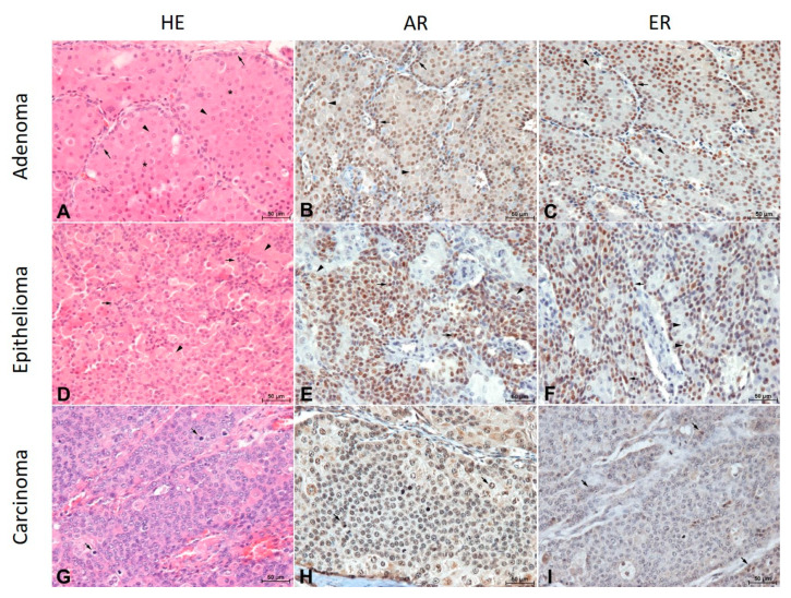
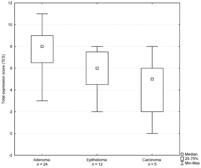
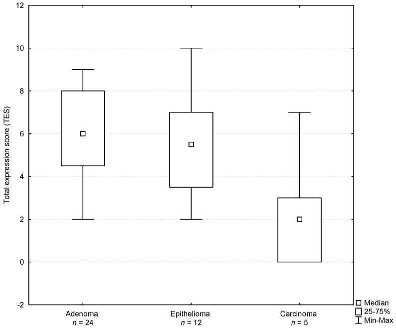
Perianal gland tumors are modified sebaceous glands present in the skin of the perianal region in the dog. Hormonal stimulation may induce hyperplasia of the perianal glands or their neoplastic progression. The presence of androgen (AR) and estrogen (ER) receptors have been demonstrated both in normal perianal glands as well as in perianal tumors. The aim of the study was an immunohistochemical assessment of the expression of estrogen and androgen receptors in perianal gland tumors in dogs as an applicatory marker for antihormonal treatment. Biopsy samples of perianal masses were collected from 41 male dogs. A histopathological examination revealed 24 adenomas, 12 epitheliomas and five carcinomas. The immunohistochemical staining showed a mainly nuclear expression of AR and ER in the neoplastic cells. Both the androgen and estrogen receptors were expressed in adenoma, epithelioma and carcinoma cases; however, the highest expression of the receptors was stated in the adenoma and epithelioma. In the case of the carcinoma, the expression of sex hormone receptors was very weak. The differences of the number of cells expressing AR and ER as well as the observed differentiated intensity of staining in the studies demonstrated that the determination of the expression of the sex hormone receptors may be useful to elaborate a diagnostic and therapeutic algorithm.

Keywords: androgen receptor; estrogen receptor; immunohistochemistry; male dogs; perianal gland tumors.


Conflict of interest statement

The authors declare no conflict of interest.

Figures

Figure 1
Figure 1
(A). Perianal gland adenoma composed of uniform broad trabeculae and islands (asterisks) of polygonal cells (arrows) surrounded by a single layer of attenuated basaloid reserve cells (arrowheads). HE, magnification × 200, bar = 50 µm. (B). Intense nuclear reaction to the androgen receptor in basaloid cells (arrows) and slightly weaker reaction in hepatoid cells (arrowheads) in a perianal gland adenoma. Indirect immunohistochemistry. Mayer’s hematoxylin counterstain, magnification × 200, bar = 50 µm. (C). A strong reaction to the estrogen receptor in basaloid cells (arrows) and a moderate reaction in hepatoid cells (arrowheads) in a perianal gland adenoma. Indirect immunohistochemistry. Mayer’s hematoxylin counterstain, magnification × 200, bar = 50 µm. (D). Proliferating basaloid cells (arrows) in the vicinity of less numerous hepatoid cells (arrowheads) in a perianal gland epithelioma. Indirect immunohistochemistry. Mayer’s hematoxylin counterstain, magnification × 200, bar = 50 µm. (E). The diversity of an androgen receptor expression in perianal gland epithelioma cells. Basaloid tumor cells were highly expressed (arrows) while hepatoid cells were predominantly low expressed (arrowheads). Indirect immunohistochemistry. Mayer’s hematoxylin counterstain, magnification × 200, bar = 50 µm. (F). A strong nuclear estrogen receptor expression in proliferating basaloid cells (arrows) and moderate to low expression in hepatoid cells (arrowheads) in a perianal gland epithelioma. Indirect immunohistochemistry. Mayer’s hematoxylin counterstain, magnification × 200, bar = 50 µm. (G). Perianal gland carcinoma. Undifferentiated, pleomorphic cells with numerous hyperchromatic cell nuclei and mitotic figures (arrows). HE, magnification × 200, bar = 50 µm. (H). Low-differentiated cells of a perianal gland carcinoma, some with mitotic figures (arrows) with a low expression of the androgen receptor. Indirect immunohistochemistry. Mayer’s hematoxylin counterstain, magnification × 200, bar = 50 µm. (I). A low expression of the estrogen receptor in perianal gland carcinoma cells (arrows). Indirect immunohistochemistry. Mayer’s hematoxylin counterstain, magnification × 200, bar = 50 µm.
Figure 2
Figure 2
Total expression score determined for androgen receptors in perianal gland tumors of male dogs.
Figure 3
Figure 3
Total expression score determined for estrogen receptors in perianal gland tumors of male dogs.

Similar articles

Cited by

References

    1. Turek M.M., Withrow S.J. Perianal tumors. In: Withrow S.J., MacEwen E.G., Page R.L., editors. Small Animal Clinical Oncolog. 5th ed. Elsevier Saunders; St. Louis, MO, USA: 2013. pp. 423–431.
    1. Brodzki A., Sobczyńska-Rak A., Brodzki P., Tatara M.R., Silmanowicz P. Occurrence, etiology and antihormonal treatment of perianal gland tumors in male dogs. Med. Weter. 2014;70:638–643.
    1. Maita K., Ishida K. Structure and development of the perianal gland of the dog. Jpn. J. Vet. Sci. 1975;37:349–356. doi: 10.1292/jvms1939.37.349. - DOI - PubMed
    1. Isitor G.N., Weinman D.E. Origin and early development of canine circumanal glands. Am. J. Vet. Res. 1979;40:487–492. - PubMed
    1. Hayes H.M., Jr., Wilson G.P. Hormone-dependent neoplasms of the canine perianal gland. Cancer Res. 1977;37:2068–2071. - PubMed

LinkOut - more resources