Review
. 2021 Mar 19;13(6):1396.
doi: 10.3390/cancers13061396.

Imaging and Management of Bladder Cancer

Affiliations
Review

Imaging and Management of Bladder Cancer

Vincenzo K Wong et al. Cancers (Basel). .

Abstract

Methods: Keyword searches of Medline, PubMed, and the Cochrane Library for manuscripts published in English, and searches of references cited in selected articles to identify additional relevant papers. Abstracts sponsored by various societies including the American Urological Association (AUA), European Association of Urology (EAU), and European Society for Medical Oncology (ESMO) were also searched.

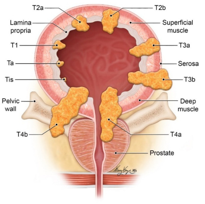
Background: Bladder cancer is the sixth most common cancer in the United States, and one of the most expensive in terms of cancer care. The overwhelming majority are urothelial carcinomas, more often non-muscle invasive rather than muscle-invasive. Bladder cancer is usually diagnosed after work up for hematuria. While the workup for gross hematuria remains CT urography and cystoscopy, the workup for microscopic hematuria was recently updated in 2020 by the American Urologic Association with a more risk-based approach. Bladder cancer is confirmed and staged by transurethral resection of bladder tumor. One of the main goals in staging is determining the presence or absence of muscle invasion by tumor which has wide implications in regards to management and prognosis. CT urography is the main imaging technique in the workup of bladder cancer. There is growing interest in advanced imaging techniques such as multiparametric MRI for local staging, as well as standardized imaging and reporting system with the recently created Vesicle Imaging Reporting and Data System (VI-RADS). Therapies for bladder cancer are rapidly evolving with immune checkpoint inhibitors, particularly programmed death ligand 1 (PD-L1) and programmed cell death protein 1 (PD-1) inhibitors, as well as another class of immunotherapy called an antibody-drug conjugate which consists of a cytotoxic drug conjugated to monoclonal antibodies against a specific target.

Conclusion: Bladder cancer is a complex disease, and its management is evolving. Advances in therapy, understanding of the disease, and advanced imaging have ushered in a period of rapid change in the care of bladder cancer patients.

Keywords: CT; MRI; bladder cancer; imaging; management; urography; urothelial carcinoma.


Conflict of interest statement

The authors declare no conflict of interest.

Figures

Figure 1
Figure 1
Diagram illustrating the different T-stages of bladder cancer. (© 2021 The University of Texas MD Anderson Cancer Center).
Figure 2
Figure 2
CT urogram showing synchronous upper and lower tract urothelial carcinoma. (a) Coronal CT image in the excretory phase shows multiple small bladder lesions (solid white arrow) and synchronous upper tract disease (dashed white arrow) (b) Coronal CT image slightly posteriorly, showing multiple upper tract lesions. (c) Coronal 3D volume rendering redemonstrates the findings with another bladder lesion (black arrow).
Figure 3
Figure 3
CT of a bladder mass during the urothelial phase and excretory phase (a) Axial CT image obtained during the urothelial phase shows a hyperenhancing bladder mass. Bladder tumors tend to be hypervascular and scanning the pelvis during the urothelial phase on CT may aid in tumor evaluation. (b) Axial CT image obtained during the excretory phase shows the same mass as a filling defect surrounded by excreted urine in the bladder.
Figure 4
Figure 4
CT urogram with partially calcified urothelial carcinoma within a large bladder diverticulum (a) Axial CT image obtained without contrast shows a large bladder diverticulum containing a partially calcified lesion along the posterior aspect representing urothelial carcinoma. (b) Axial CT image obtained during the excretory phase and (c) Sagittal excretory phase image show the same mass as a filling defect surrounded by excreted urine in the bladder.
Figure 5
Figure 5
Muscle invasive bladder cancer on MRI (a) Axial T2WI shows a muscle invasive mass in the right bladder wall, seen as a T2 intermediate signal lesion (solid white arrow) invasive into the muscularis propria which is T2 hypointense. The mass is located at the ureterovesicular junction, causing hydroureter (dashed white arrow). (b) Axial DWI image, showing the hyperintense mass invading the muscularis propria, which is T2 intermediate signal on DWI. (c) ADC map showing hypointensity in the area of DWI hyperintensity, confirming restricted diffusion.
Figure 6
Figure 6
MR urography obtained in a patient with Lynch syndrome showing synchronous upper and lower tract lesions. Pathology of the bladder lesion confirmed non-invasive urothelial carcinoma and upper tract washings were concerning for low grade urothelial carcinoma. (a) Coronal post-contrast T1WI shows enhancing lesions in the bladder (solid white arrow) and left renal pelvis (dashed white arrow). (b) Coronal post-contrast excretory phase image shows the lesions as filling defects, surrounded by excreted contrast. (c) Coronal T2WI shows the upper tract lesion surrounded by T2 hyperintense urine. (d) Coronal T2WI of the bladder shows the bladder lesion as T2 intermediate in signal without invasion into the T2 hypointense muscularis propria. (e) Axial T2WI of the bladder shows the mass to be non-invasive. (f) Axial DWI shows restricted diffusion of the mass.
Figure 7
Figure 7
Marked progression of locally advanced bladder cancer and metastatic disease. (a) Axial CT image demonstrates a small lesion in the anterior bladder which went untreated for 16 months. (b) Follow up axial CT image showing significant progression of the mass which now extends beyond the bladder to involve the uterus, right adnexa, and right pelvic side wall. (c) Axial PET/CT image redemonstrating the locally advanced mass with bilateral pelvic lymphadenopathy. (d) Axial PET/CT image shows metastatic retroperitoneal lymphadenopathy. (e) Coronal PET/CT image also showing metastatic left supraclavicular lymphadenopathy. Biopsy of the left supraclavicular node showed poorly differentiated carcinoma compatible with bladder primary.
Figure 7
Figure 7
Marked progression of locally advanced bladder cancer and metastatic disease. (a) Axial CT image demonstrates a small lesion in the anterior bladder which went untreated for 16 months. (b) Follow up axial CT image showing significant progression of the mass which now extends beyond the bladder to involve the uterus, right adnexa, and right pelvic side wall. (c) Axial PET/CT image redemonstrating the locally advanced mass with bilateral pelvic lymphadenopathy. (d) Axial PET/CT image shows metastatic retroperitoneal lymphadenopathy. (e) Coronal PET/CT image also showing metastatic left supraclavicular lymphadenopathy. Biopsy of the left supraclavicular node showed poorly differentiated carcinoma compatible with bladder primary.
Figure 8
Figure 8
Ultrasound and CT images of non-muscle invasive bladder cancer. (a) Longitudinal grayscale ultrasound image of the bladder shows a large mass within the bladder. (b) Transverse color Doppler image shows vascularity within the mass. (c) Axial portal venous phase CT image shows a central area of enhancement, similar to the color Doppler ultrasound image. (d) Axial delayed phase CT image shows the mass partially surrounded by excreted contrast in the bladder.
Figure 9
Figure 9
IVP and CT images of locally advanced bladder cancer. (a) IVP image of the upper urinary tract at 9-min is without filling defect in the left collecting system to suggest left-sided upper tract disease. The right collecting system is poorly opacified secondary to hydroureteronephrosis, better depicted on CT. (b) IVP image at 15 min shows a large filling defect in the bladder. (c) Magnified oblique view of the bladder better show a posterior filling defect. (d) Axial portal venous phase CT image of the pelvis shows the corresponding mass in the posterior bladder which extends posteriorly to involve the vagina. (e) Coronal portal venous phase CT image shows the bladder mass causing right hydrouteronephrosis.

References

    1. Galsky M.D., Hahn N.M., Rosenberg J., Sonpavde G., Hutson T., Oh W.K., Dreicer R., Vogelzang N., Sternberg C.N., Bajorin D.F., et al. Treatment of patients with metastatic urothelial cancer “unfit” for Cisplatin-based chemotherapy. J. Clin. Oncol. 2011;29:2432–2438. doi: 10.1200/JCO.2011.34.8433. - DOI - PubMed
    1. Sloan F.A., Yashkin A.P., Akushevich I., Inman B.A. The Cost to Medicare of Bladder Cancer Care. Eur. Urol. Oncol. 2020;3:515–522. doi: 10.1016/j.euo.2019.01.015. - DOI - PubMed
    1. Al-Husseini M.J., Kunbaz A., Saad A.M., Santos J.V., Salahia S., Iqbal M., Alahdab F. Trends in the incidence and mortality of transitional cell carcinoma of the bladder for the last four decades in the USA: A SEER-based analysis. BMC Cancer. 2019;19:46. doi: 10.1186/s12885-019-5267-3. - DOI - PMC - PubMed
    1. Humphrey P.A., Moch H., Cubilla A.L., Ulbright T.M., Reuter V.E. The 2016 WHO Classification of Tumours of the Urinary System and Male Genital Organs-Part B: Prostate and Bladder Tumours. Eur. Urol. 2016;70:106–119. doi: 10.1016/j.eururo.2016.02.028. - DOI - PubMed
    1. Willis D., Kamat A.M. Nonurothelial bladder cancer and rare variant histologies. Hematol. Oncol. Clin. N. Am. 2015;29:237–252. doi: 10.1016/j.hoc.2014.10.011. - DOI - PubMed

LinkOut - more resources