. 2021 Mar 19;10(3):681.
doi: 10.3390/cells10030681.

Metformin Attenuates Monosodium-Iodoacetate-Induced Osteoarthritis via Regulation of Pain Mediators and the Autophagy-Lysosomal Pathway

Affiliations

Metformin Attenuates Monosodium-Iodoacetate-Induced Osteoarthritis via Regulation of Pain Mediators and the Autophagy-Lysosomal Pathway

Hyun Sik Na et al. Cells. .

Abstract

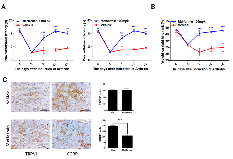
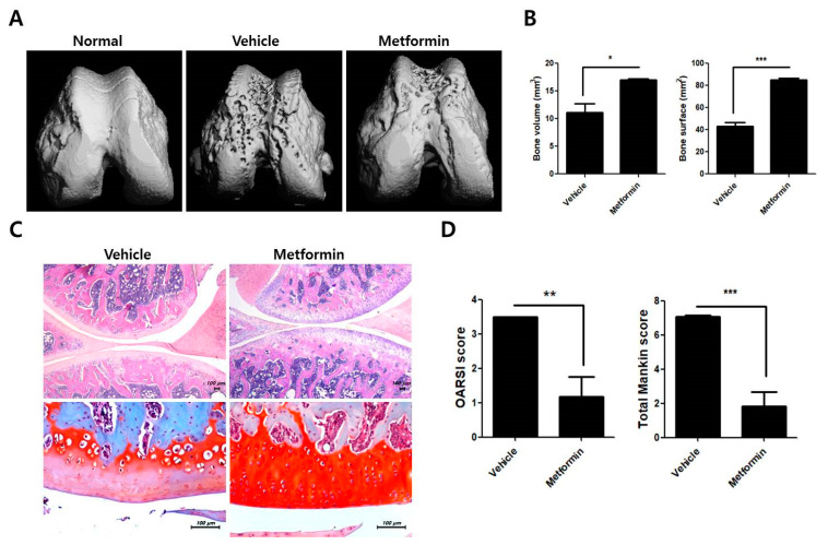
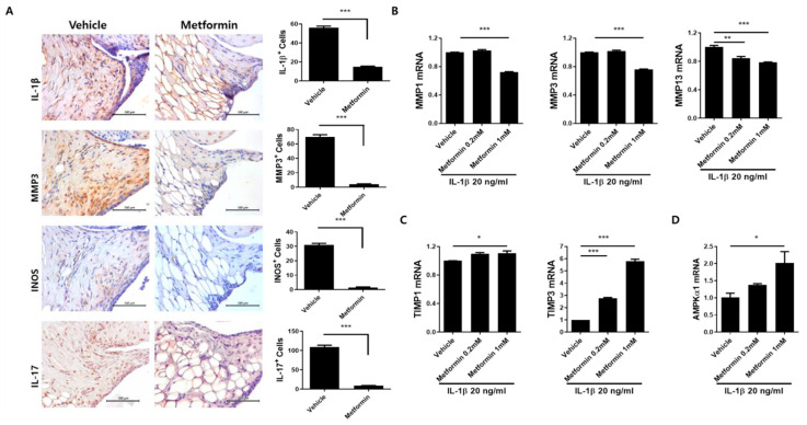
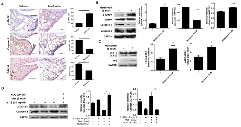
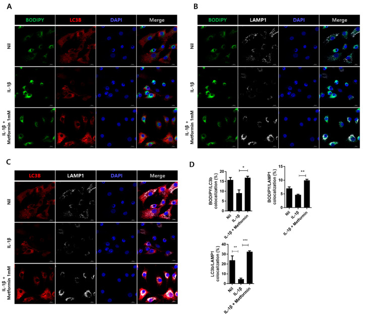
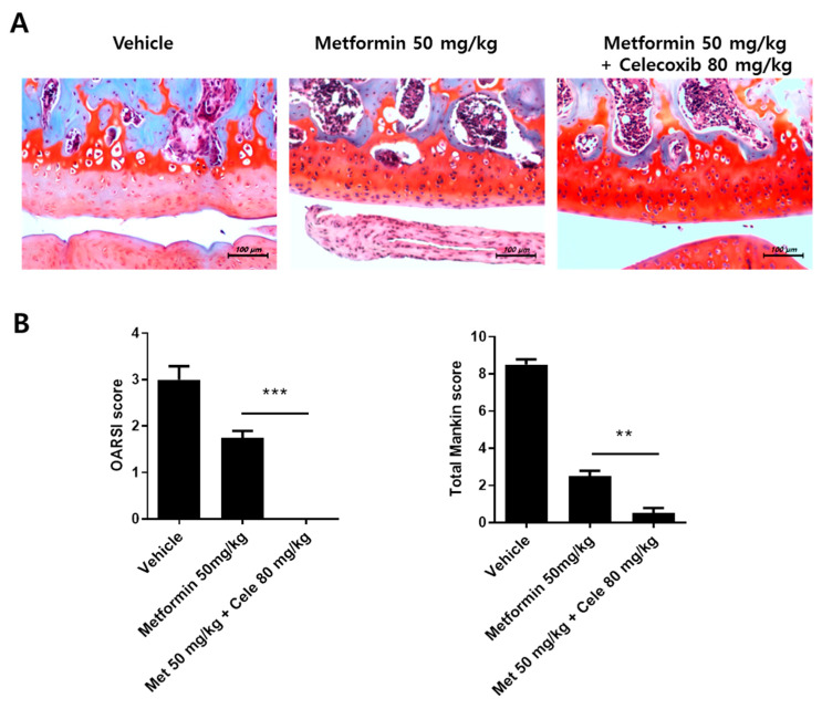
Osteoarthritis (OA) is the most common degenerative arthritis associated with pain and cartilage destruction in the elderly; it is known to be involved in inflammation as well. A drug called celecoxib is commonly used in patients with osteoarthritis to control pain. Metformin is used to treat type 2 diabetes but also exhibits regulation of the autophagy pathway. The purpose of this study is to investigate whether metformin can treat monosodium iodoacetate (MIA)-induced OA in rats. Metformin was administered orally every day to rats with OA. Paw-withdrawal latency and threshold were used to assess pain severity. Cartilage damage and pain mediators in dorsal root ganglia were evaluated by histological analysis and a scoring system. Relative mRNA expression was measured by real-time PCR. Metformin reduced the progression of experimental OA and showed both antinociceptive properties and cartilage protection. The combined administration of metformin and celecoxib controlled cartilage damage more effectively than metformin alone. In chondrocytes from OA patients, metformin reduced catabolic factor gene expression and inflammatory cell death factor expression, increased LC3Ⅱb, p62, and LAMP1 expression, and induced an autophagy-lysosome fusion phenotype. We investigated if metformin treatment reduces cartilage damage and inflammatory cell death of chondrocytes. The results suggest the potential for the therapeutic use of metformin in OA patients based on its ability to suppress pain and protect cartilage.

Keywords: autophagy; cartilage; combination therapy; metformin; osteoarthritis; pain.


Conflict of interest statement

The authors declare that they have no competing interest.

Figures

Figure 1
Figure 1
Metformin reduces pain in monosodium iodoacetate (MIA)-induced OA rats. The rats were injected with 3 mg of MIA. After osteoarthritis (OA) induction, the rats were administrated with metformin 100 mg/kg (N = 6 animals per group). (A) Nociceptive testing was performed using a dynamic plantar aesthesiometer (Ugo Basile, Gemonio, Italy), an automated version of the von Frey hair assessment procedure. (B) Weight-bearing was assessed using an incapacitance meter (IITC Life Science, Woodland Hills, CA, USA). (C) Dorsal root ganglion tissue from rats in all groups was stained immunohistochemically with specific antibodies for CGRP and TRPV1. Immunohistochemically identified TRPV1- and CGRP-positive cells were counted. Data are represented as the mean ± standard error of the mean (SEM) of three independent experiments (*** p < 0.001).
Figure 2
Figure 2
Metformin ameliorates bone and cartilage erosion in MIA-induced OA rats. The rats were injected with 3 mg of MIA. After OA induction, the rats were administrated with metformin 100 mg/kg (N = 6 animals per group). The rats were sacrificed at Day 15 after OA induction to collect joint tissues. (A) Twenty cylindrical bone samples were obtained from bone biopsies of 20 dry hemimandibles. The samples of six animals per group were scanned using microcomputed tomography (mCT 35; SCANCO Medical, Zurich, Switzerland). (B) Bone volume and bone surface were analyzed using NRecon software. (C) Knee-joint tissue samples were acquired from all OA groups at 2 weeks and stained with hematoxylin and eosin (H&E) and safranin O (D) to determine Osteoarthritis Research Society International (OARSI) and Mankin scores. Data are represented as the mean ± SEM of three independent experiments (* p < 0.05, ** p < 0.01, *** p < 0.001).
Figure 3
Figure 3
Expression of Inflammatory mediators following metformin administration. The rats were injected with 3 mg of MIA. After OA induction, the rats were administrated with metformin 100 mg/kg (N = 6 animals per group). The OA rats were sacrificed at Day 15 after OA induction to collect joint tissues. (A) The expression of IL-1β, MMP3, iNOS, and IL-17 after metformin administration was determined immunohistochemically in the synovium of rats with MIA-induced OA. The count of positive cells for each antibody is shown on the right. The data are reported as the mean ± SD of three independent experiments, with six animals per group. *** p < 0.001. (B) The human OA chondrocytes were cultured with metformin (0.2 and 1 mM) and IL-1β (20 ng/mL) for 24 h. mRNA levels for MMP-1, -3, and -13, (C) TIMP-1, -3, and (D) AMPKα1 were measured by real-time-PCR. Data are represented as the mean ± SEM of three independent experiments (* p < 0.05, ** p < 0.01, *** p < 0.001).
Figure 4
Figure 4
Expression of cell death mediators following metformin administration. (A) The expression of p-AMPK, caspase-1, and p-MLKL in the knee joint tissues of MIA-induced OA rats was determined immunohistochemically (N = 6 animals per group). The count of positive cells for each antibody is shown on the right. Human OA chondrocytes were cultured with metformin (5 mM) for 24 h. (B,C) p-AMPK, caspase-1, caspase-3, LC3, p62, and GAPDH were analyzed using Western blot in the protein of metformin-treated human chondrocytes. (D) The human OA chondrocytes were cultured with metformin (5 mM), IL-1β (20 ng/mL), and HCQ (20 μM) for 24 h. Caspase-1, caspase-3, and GAPDH were analyzed using Western blot. Data are represented as the mean ± SEM of three independent experiments (* p < 0.05, ** p < 0.01, *** p < 0.001).
Figure 5
Figure 5
Metformin regulates autophagolysosome via LAMP1 activation. Human chondrocytes were cultured with metformin (1 mM) and IL-1β (20 ng/mL) for 24 h. The cells were analyzed using confocal microscopy for autophagosome and autophagolysosome. For autophagosome structure analysis, (A) colocalization of BODIPY (green) and LC3B (red) were analyzed in unstimulated (Nil) or each stimulated human chondrocyte (B,C) for autophagolysosome, colocalization of BODIPY (green), and LAMP1 (white) or colocalization of LC3B (red) and LAMP1 (white). (D) Colocalization fluorescence was analyzed using the Fiji/ImageJ program. The data are a repeat of three independent experiments, and the fluorescent data are presented as the mean ± SEM of three independent experiments (* p < 0.05, ** p < 0.01, *** p < 0.001).
Figure 6
Figure 6
Combined effects of metformin and celecoxib. (A) OA was induced in Wistar rats by intra-articular injection of MIA. Metformin (50 mg/kg) and celecoxib (80 mg/kg) were orally administrated into OA rats for 14 days (N = 6 animals per group). Knee-joint tissue samples acquired from all OA groups at 14 days were stained with H&E and safranin O (B) to evaluate disease severity based on OARSI and Mankin scores. Data are represented as the mean ± SEM of three independent experiments (** p < 0.01, *** p < 0.001).
Figure 7
Figure 7
Clinical study of the effect of metformin in OA patients. A clinical assessment of OA patients with type 2 diabetes, who had been followed-up for >3 years, showed slower progression of OA in those taking metformin (nondiabetic = 23 patients, diabetic with metformin = 18 patients; all patients female). The OA grade was analyzed based on 3 years of data (* p < 0.05).

References

    1. Kalman D.S., Heimer M., Valdeon A., Schwartz H., Sheldon E. Effect of a natural extract of chicken combs with a high content of hyaluronic acid (Hyal-Joint) on pain relief and quality of life in subjects with knee osteoarthritis: A pilot randomized double-blind placebo-controlled trial. Nutr. J. 2008;7:3. doi: 10.1186/1475-2891-7-3. - DOI - PMC - PubMed
    1. Tonge D.P., Pearson M.J., Jones S.W. The hallmarks of osteoarthritis and the potential to develop personalised disease-modifying pharmacological therapeutics. Osteoarthr. Cart. 2014;22:609–621. doi: 10.1016/j.joca.2014.03.004. - DOI - PubMed
    1. Berenbaum F. Osteoarthritis as an inflammatory disease (osteoarthritis is not osteoarthrosis!) Osteoarthr. Cart. 2013;21:16–21. doi: 10.1016/j.joca.2012.11.012. - DOI - PubMed
    1. Kapoor M., Martel-Pelletier J., Lajeunesse D., Pelletier J.P., Fahmi H. Role of proinflammatory cytokines in the pathophysiology of osteoarthritis. Nat. Rev. Rheumatol. 2011;7:33–42. doi: 10.1038/nrrheum.2010.196. - DOI - PubMed
    1. Sokolove J., Lepus C.M. Role of inflammation in the pathogenesis of osteoarthritis: Latest findings and interpretations. Adv. Musculoskelet Dis. 2013;5:77–94. doi: 10.1177/1759720X12467868. - DOI - PMC - PubMed

Publication types

LinkOut - more resources