. 2021 Mar 19;10(3):682.
doi: 10.3390/cells10030682.

Graphene Oxide-Silver Nanoparticle Nanocomposites Induce Oxidative Stress and Aberrant Methylation in Caprine Fetal Fibroblast Cells

Affiliations

Graphene Oxide-Silver Nanoparticle Nanocomposites Induce Oxidative Stress and Aberrant Methylation in Caprine Fetal Fibroblast Cells

Yu-Guo Yuan et al. Cells. .

Abstract

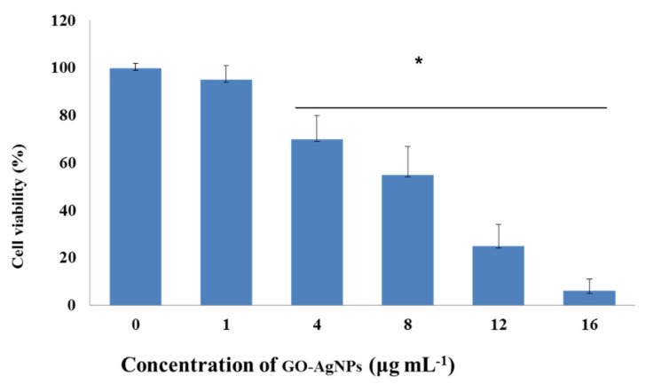
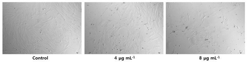
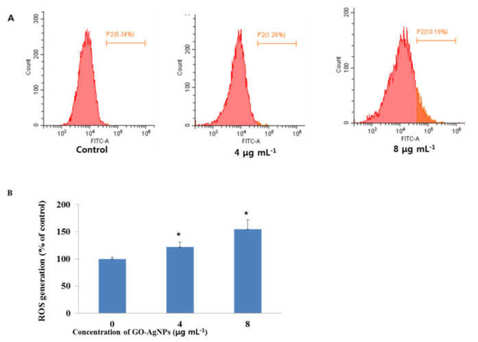
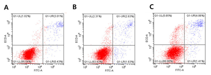
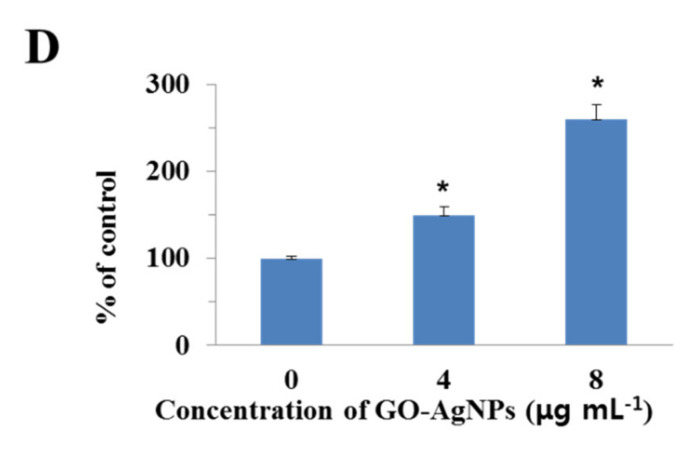
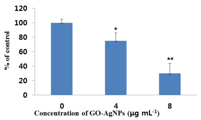
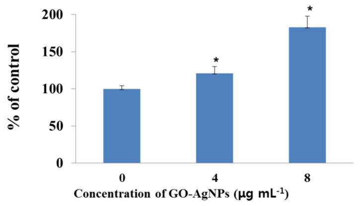
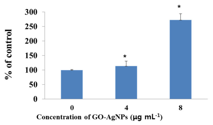
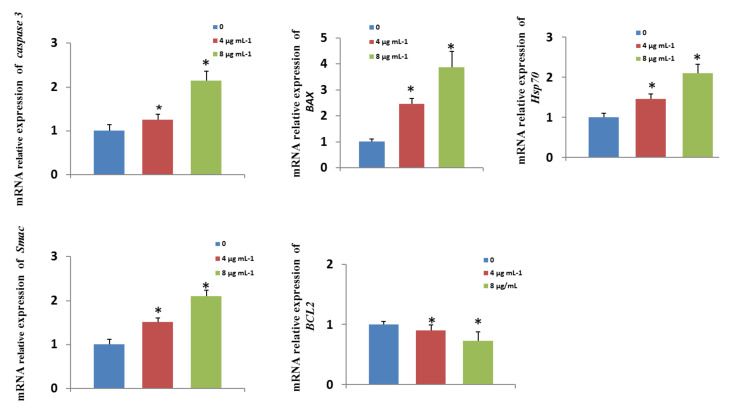
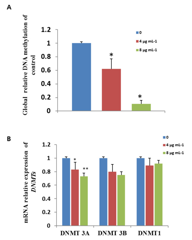
Graphene oxide-silver nanoparticle (GO-AgNPs) nanocomposites have drawn much attention for their potential in biomedical uses. However, the potential toxicity of GO-AgNPs in animals and humans remains unknown, particularly in the developing fetus. Here, we reported the GO-AgNP-mediated cytotoxicity and epigenetic alteration status in caprine fetal fibroblast cells (CFFCs). In brief, the proliferation and apoptosis rate of GO-AgNP-treated CFFCs (4 and 8 µg/mL of GO-AgNPs) were measured using the cell-counting kit (CCK-8) assay and the annexin V/propidium iodide (PI) assay, respectively. In addition, the oxidative stress induced by GO-AgNPs and detailed mechanisms were studied by evaluating the generation of reactive oxygen species (ROS), superoxide dismutase (SOD), lactate dehydrogenase (LDH), malondialdehyde (MDA), and caspase-3 and abnormal methylation. The expression of pro- and anti-apoptotic genes and DNA methyltransferases was measured using reverse transcription followed by RT-qPCR. Our data indicated that GO-AgNPs cause cytotoxicity in a dose-dependent manner. GO-AgNPs induced significant cytotoxicity by the loss of cell viability, production of ROS, increasing leakage of LDH and level of MDA, increasing expression of pro-apoptotic genes, and decreasing expression of anti-apoptotic genes. GO-AgNPs incited DNA hypomethylation and the decreased expression of DNMT3A. Taken together, this study showed that GO-AgNPs increase the generation of ROS and cause apoptosis and DNA hypomethylation in CFFCs. Therefore, the potential applications of GO-AgNPs in biomedicine should be re-evaluated.

Keywords: caprine fetal fibroblast cells (CFFCs); epigenetic; graphene oxide; reactive oxygen species (ROS); silver nanoparticles.


Conflict of interest statement

The authors declare no conflict of interest.

Figures

Figure 1
Figure 1
Size determination analysis of graphene oxide–silver nanoparticle (GO-AgNP) nanocomposites by transmission electron microscopy (TEM). The TEM image showed that the size of GO-AgNPs was about 20 nm.
Figure 2
Figure 2
Effects of GO-AgNPs on the proliferation of caprine fetal fibroblast cells (CFFCs). CFFCs were exposed to 0, 1, 4, 8, 12, and 16 µg/mL of GO-AgNPs for 24 h. The percentage of cell viability was then calculated relative to the control group (0 µg/mL). Values are presented as the mean ± SD of four independent experiments (* p < 0.05).
Figure 3
Figure 3
Cell morphology following treatment with GO-AgNPs. Caprine fetal fibroblast cells (CFFCs) were treated with 0, 4, and 8 µg/mL of GO-AgNPs for 24 h and then visualized under a phase-contrast microscope (magnification, 100×). Scale bar = 20 μm.
Figure 4
Figure 4
Total reactive oxygen species (ROS) generation in GO-AgNP-treated cells. Caprine fetal fibroblast cells (CFFCs) were treated with 0, 4, and 8 µg/mL of GO-AgNPs for 24 h and analyzed by FACS (A). The percentage of ROS generation relative to the untreated control group (0 µg/mL) (B). Values are presented as the mean ± SD of four independent experiments (* p < 0.05).
Figure 5
Figure 5
Evaluation of GO-AgNP-induced apoptotic cell death with the annexin V–FITC/propidium iodide (PI) staining assay. Caprine fetal fibroblast cells (CFFCs) were treated with 0 (A), 4 (B), and 8 µg/mL (C) of GO-AgNPs for 24 h, and FACS was carried out for detection of fractions of early apoptotic, late apoptotic, and necrotic CFFCs. The corresponding linear diagram of flow cytometry is shown in (D). Values are presented as the mean ± SD of five independent experiments (* p < 0.05).
Figure 5
Figure 5
Evaluation of GO-AgNP-induced apoptotic cell death with the annexin V–FITC/propidium iodide (PI) staining assay. Caprine fetal fibroblast cells (CFFCs) were treated with 0 (A), 4 (B), and 8 µg/mL (C) of GO-AgNPs for 24 h, and FACS was carried out for detection of fractions of early apoptotic, late apoptotic, and necrotic CFFCs. The corresponding linear diagram of flow cytometry is shown in (D). Values are presented as the mean ± SD of five independent experiments (* p < 0.05).
Figure 6
Figure 6
Measurement of superoxide dismutase (SOD) production in GO-AgNP-treated cells. Caprine fetal fibroblast cells (CFFCs) were treated with 0, 4, and 8 µg/mL of GO-AgNPs for 24 h. The percentage of SOD relative to the control group (0 µg/mL) was determined. Values are presented as the mean ± SD of four independent experiments (* p < 0.05; ** p < 0.01).
Figure 7
Figure 7
Measurement of malondialdehyde (MDA) production in GO-AgNP-treated cells. Caprine fetal fibroblast cells (CFFCs) were treated with 0, 4, and 8 µg/mL of GO-AgNPs for 24 h. The percentage of MDA relative to the control group (0 µg/mL) was determined. Values are presented as the mean ± SD of four independent experiments (* p < 0.05).
Figure 8
Figure 8
Measurement of lactate dehydrogenase (LDH) activity in GO-AgNP-treated cells. Caprine fetal fibroblast cells (CFFCs) were treated with 0, 4, and 8 µg/mL of GO-AgNPs for 24 h. The percentage of LDH activity relative to the control group (0 µg/mL) was determined. Values are presented as the mean ± SD of five independent experiments (* p < 0.05).
Figure 9
Figure 9
Measurement of caspase-3 activity in GO-AgNP-treated cells. Caprine fetal fibroblast cells (CFFCs) were treated with 0, 4, and 8 µg/mL of GO-AgNPs for 24 h. The percentage of caspase-3 activity relative to the control group (0 µg/mL) was determined. Values are presented as the mean ± SD of four independent experiments (* p < 0.05).
Figure 10
Figure 10
Effects of GO-AgNPs on apoptotic gene expression levels. Caprine fetal fibroblast cells (CFFCs) were treated with 0, 4, and 8 µg/mL of GO-AgNPs for 24 h. Relative mRNA levels of genes related to apoptosis were determined. Values are presented as the mean ± SD of four independent experiments (* p < 0.05).
Figure 11
Figure 11
Effects of GO-AgNPs on global DNA methylation and gene expression levels. Caprine fetal fibroblast cells (CFFCs) were treated with 0, 4, and 8 µg/mL of GO-AgNPs for 24 h. Relative global DNA methylation (A) and mRNA levels of DNMTs (B) were determined. Values are presented as the mean ± SD of four independent experiments (* p < 0.05; ** p < 0.01).

Similar articles

Cited by

References

    1. Pinho A.R., Martins F., Costa M., Senos A., Silva O., Pereira M.L., Rebelo S. In vitro cytotoxicity effects of zinc oxide nanoparticles on spermatogonia cells. Cells. 2020;9:1081. doi: 10.3390/cells9051081. - DOI - PMC - PubMed
    1. Gurunathan S., Kim J.H. Graphene oxide-silver nanoparticles nanocomposite stimulates differentiation in human neuroblastoma cancer cells (SH-SY5Y) Int. J. Mol. Sci. 2017;18:2549. doi: 10.3390/ijms18122549. - DOI - PMC - PubMed
    1. Gies V., Zou S. Systematic toxicity investigation of graphene oxide: Evaluation of assay selection, cell type, exposure period and flake size. Toxicol. Res. 2018;7:93–101. doi: 10.1039/C7TX00278E. - DOI - PMC - PubMed
    1. Gurunathan S., Han J.W., Eppakayala V., Dayem A.A., Kwon D.N., Kim J.H. Biocompatibility effects of biologically synthesized graphene in primary mouse embryonic fibroblast cells. Nanoscale. Res. Lett. 2013;8:393. doi: 10.1186/1556-276X-8-393. - DOI - PMC - PubMed
    1. Thapa R.K., Kim J.H., Jeong J.H., Shin B.S., Choi H.G., Yong C.S., Kim J.O. Silver nanoparticle-embedded graphene oxide-methotrexate for targeted cancer treatment. Colloids. Surf. B. Biointerfaces. 2017;153:95–103. doi: 10.1016/j.colsurfb.2017.02.012. - DOI - PubMed

Publication types

LinkOut - more resources