Review
. 2021 Mar 19;11(3):789.
doi: 10.3390/nano11030789.

Novel Inorganic Nanomaterial-Based Therapy for Bone Tissue Regeneration

Affiliations
Review

Novel Inorganic Nanomaterial-Based Therapy for Bone Tissue Regeneration

Yu Fu et al. Nanomaterials (Basel). .

Abstract

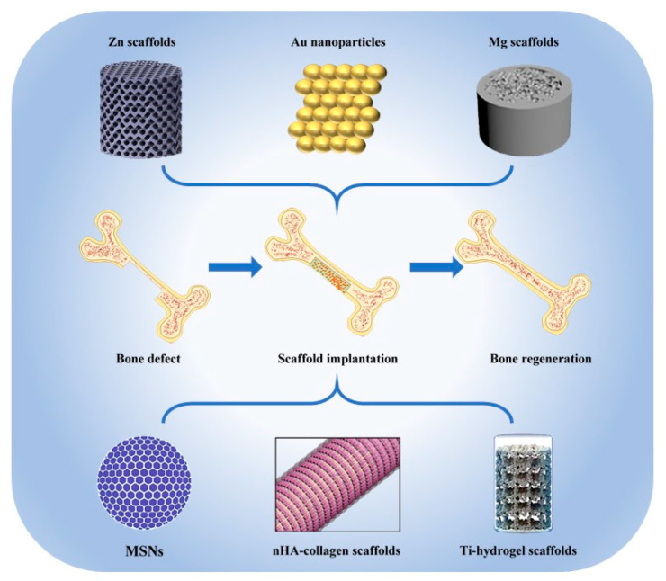
Extensive bone defect repair remains a clinical challenge, since ideal implantable scaffolds require the integration of excellent biocompatibility, sufficient mechanical strength and high biological activity to support bone regeneration. The inorganic nanomaterial-based therapy is of great significance due to their excellent mechanical properties, adjustable biological interface and diversified functions. Calcium-phosphorus compounds, silica and metal-based materials are the most common categories of inorganic nanomaterials for bone defect repairing. Nano hydroxyapatites, similar to natural bone apatite minerals in terms of physiochemical and biological activities, are the most widely studied in the field of biomineralization. Nano silica could realize the bone-like hierarchical structure through biosilica mineralization process, and biomimetic silicifications could stimulate osteoblast activity for bone formation and also inhibit osteoclast differentiation. Novel metallic nanomaterials, including Ti, Mg, Zn and alloys, possess remarkable strength and stress absorption capacity, which could overcome the drawbacks of low mechanical properties of polymer-based materials and the brittleness of bioceramics. Moreover, the biodegradability, antibacterial activity and stem cell inducibility of metal nanomaterials can promote bone regeneration. In this review, the advantages of the novel inorganic nanomaterial-based therapy are summarized, laying the foundation for the development of novel bone regeneration strategies in future.

Keywords: bone regeneration; inorganic nanomaterials; metallic nanomaterials; nano hydroxyapatites; nano silica.


Conflict of interest statement

The authors declare no conflict of interest.

Figures

Figure 1
Figure 1
The main inorganic nanomaterials used for bone tissue regeneration [10,11,12,13,14].
Figure 2
Figure 2
The mechanical and biological properties of nHA/polymer composites. (a) The highly ordered deposited nano hydroxyapatites (nHAs) provided intrafibrillarly mineralized collagen (IMC) with much higher Young’s modulus than that of pure collagen (COL) and extrafibrillarly mineralized collagen (EMC) [16]. (b) The addition of nHAs increased the compressive modulus of silk fibroin significantly [22]. (c) nHA/kappa-carrageenan (κ-CG) enhanced the alkaline phosphatase (ALP) activity and calcium deposition of human osteoblasts, regardless of the concentration of κ-CG [28]. (d) With the increase of nHAs content, the MC3T3-E1 preosteoblast cells expressed more osteocalcin (OCN) and bone sialoprotein (BSP) [24]. (e) The nHA/silk fibroin composites promoted the bone regeneration in rat calvarial defects [22]. The co-inspired scaffold (CIS) which consisted of nHAs and chitosan induced both new bone formation (f) and vascularization (g) compared with blank and pure chitosan [29]. *: p < 0.05, **: p < 0.01, ***: p < 0.001.
Figure 3
Figure 3
The properties affecting bone regeneration of nHA-based 3D printing scaffolds. (a) Histomorphometric examination demonstrated that the 3D printed HA/ tricalcium phosphate scaffold (3DS) had better bone reparation effect than both negative control (NS) and positive control (particle-type bone substitutes, PS) in beagle dog mandibular bone defects [30]. (b) Radiographic analysis showed that 3D hydroxyapatite/extracellular matrix (HA/ECM) composites promoted bone regeneration in rat calvarial defects after 12 weeks [34]. (c) Both radiographic and histological evaluations exhibited more new bone formation for 3D printed nHA-based scaffolds with higher pore size only at 4 weeks [35]. *: p < 0.05, **: p < 0.01, ***: p < 0.001.
Figure 4
Figure 4
The application of nano silica in bone tissue engineering. (a) The effect of Si ions on osteogenesis-related gene expression of human bone marrow mesenchymal stem cells (BMSCs). A: blank control (DMEM); B: 10 μg/mL Si ions in DMEM; C: 50 μg/mL Si ions in DMEM; D–F: A–C solution plus 35 μg/mL dimethyloxaloylglycine, respectively. *: Comparison between blank and other groups; #: comparison between group B and E, C and F, respectively; p < 0.05 [13]. (b) Mesoporous silica nanoparticles (MSNs) assisted E2 sustained release, which prevented osteoporosis in vivo. OVX = ovariectomy; UCHRT = NaLuF4:Yb,Tm@NaLuF4@mSiO2-EDTA-E2 nanocomposites. *: p < 0.05, **: p < 0.01 [42]. (c) Nano silica incorporated collagen fibrils promoted bone regeneration in rat femoral defects. SCS = silicified collagen scaffold; NC: negative control; SCS + PD: silicified collagen scaffold plus PD098059; SCS + SB: silicified collagen scaffold plus SB203580; PD and SB are MAPK inhibitors. (i) Micro-CT scans and quantitative results. (ii) van Geison staining. Bar = 1 mm. (iii) Ponceau trichrome staining. Bar = 200 μm [52]. (d) The osteoconductivity of calcium/magnesium-doped rhBMP-2-incorporated MSN scaffold in vitro and in vivo. CMMS= calcium/magnesium-doped silica-based scaffolds. (i) rhBMP-2 increased the osteogenesis-related gene expression of rat BMSCs. (ii) CMMS/rhBMP-2 scaffold promoted bone regeneration in rabbit femoral defects as demonstrated by micro-CT analysis and histological evaluation. M: materials, B: bone, F: fibrous tissue. *: p < 0.05; #: p < 0.05 [53].
Figure 5
Figure 5
The biological properties of AM Ti-based implants. (a) Custom-made Ti-6Al-4V-ELI AM implants helped to reconstruct the skull defect of 21 patients [74]. (b) Silk fibroin and vancomycin coating promoted the survival of MC3T3-E1 cell line seeded on 3D printed Ti implants [82].(c) Silk fibroin and vancomycin coating promoted the calcium deposition of MC3T3-E1 cell line seeded on 3D printed Ti implants [82]. (d) The micropattern of Ti substrates induced osteogenesis. (i) SEM images and 3D surface profile of different micropatterns. (ii) MC3T3-E1 preosteoblasts aligned along the ridges in R3G7 micropattern. (iii) The R3G7 microgroove pattern promoted osteogenesis of MC3T3-E1 cells. (iv) and (v) The R3G7 microgroove pattern induced bone regeneration in rat skull defects [80]. *: p < 0.05; **: p < 0.01; ***: p < 0.001.

Similar articles

Cited by

References

    1. Wegst U.G., Bai H., Saiz E., Tomsia A.P., Ritchie R.O. Bioinspired structural materials. Nat. Mater. 2015;14:23–36. doi: 10.1038/nmat4089. - DOI - PubMed
    1. Roddy E., DeBaun M.R., Daoud-Gray A., Yang Y.P., Gardner M.J. Treatment of critical-sized bone defects: Clinical and tissue engineering perspectives. Eur. J. Orthop. Surg. Traumatol. 2018;28:351–362. doi: 10.1007/s00590-017-2063-0. - DOI - PubMed
    1. Griffin K.S., Davis K.M., McKinley T.O., Anglen J.O., Chu T.-M.G., Boerckel J.D., Kacena M.A. Evolution of Bone Grafting: Bone Grafts and Tissue Engineering Strategies for Vascularized Bone Regeneration. Clin. Rev. Bone Miner. Metab. 2015;13:232–244. doi: 10.1007/s12018-015-9194-9. - DOI
    1. Burk T., Del Valle J., Finn R.A., Phillips C. Maximum Quantity of Bone Available for Harvest From the Anterior Iliac Crest, Posterior Iliac Crest, and Proximal Tibia Using a Standardized Surgical Approach: A Cadaveric Study. J. Oral Maxillofac. Surg. 2016;74:2532–2548. doi: 10.1016/j.joms.2016.06.191. - DOI - PubMed
    1. Cross L.M., Thakur A., Jalili N.A., Detamore M., Gaharwar A.K. Nanoengineered biomaterials for repair and regeneration of orthopedic tissue interfaces. Acta Biomater. 2016;42:2–17. doi: 10.1016/j.actbio.2016.06.023. - DOI - PubMed

LinkOut - more resources