. 2021 Mar 12;13(6):1250.
doi: 10.3390/cancers13061250.

Single-Cell Expression Landscape of SARS-CoV-2 Receptor ACE2 and Host Proteases in Normal and Malignant Lung Tissues from Pulmonary Adenocarcinoma Patients

Affiliations

Single-Cell Expression Landscape of SARS-CoV-2 Receptor ACE2 and Host Proteases in Normal and Malignant Lung Tissues from Pulmonary Adenocarcinoma Patients

Guangchun Han et al. Cancers (Basel). .

Abstract

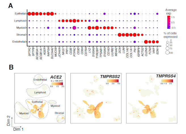
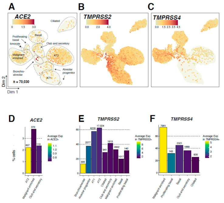
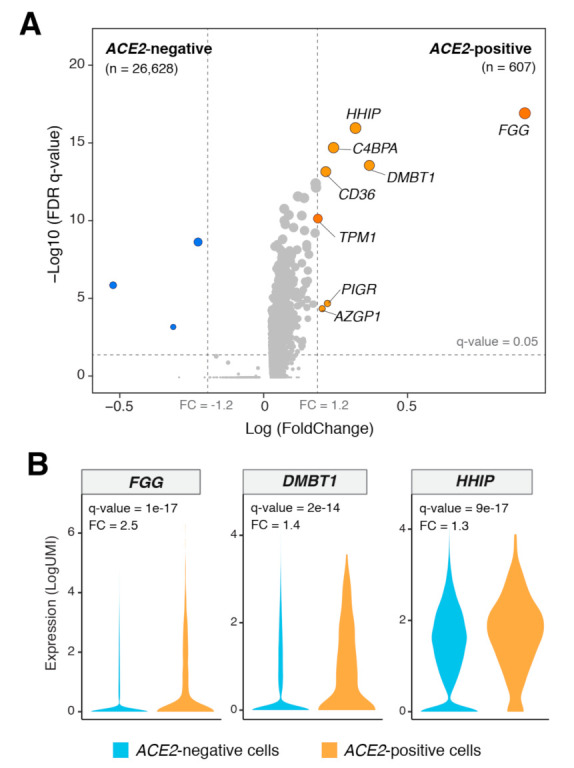
The novel coronavirus SARS-CoV-2 is the causative agent of the COVID-19 pandemic. Severely symptomatic COVID-19 is associated with lung inflammation, pneumonia, and respiratory failure, thereby raising concerns of elevated risk of COVID-19-associated mortality among lung cancer patients. Angiotensin-converting enzyme 2 (ACE2) is the major receptor for SARS-CoV-2 entry into lung cells. The single-cell expression landscape of ACE2 and other SARS-CoV-2-related genes in pulmonary tissues of lung cancer patients remains unknown. We sought to delineate single-cell expression profiles of ACE2 and other SARS-CoV-2-related genes in pulmonary tissues of lung adenocarcinoma (LUAD) patients. We examined the expression levels and cellular distribution of ACE2 and SARS-CoV-2-priming proteases TMPRSS2 and TMPRSS4 in 5 LUADs and 14 matched normal tissues by single-cell RNA-sequencing (scRNA-seq) analysis. scRNA-seq of 186,916 cells revealed epithelial-specific expression of ACE2, TMPRSS2, and TMPRSS4. Analysis of 70,030 LUAD- and normal-derived epithelial cells showed that ACE2 levels were highest in normal alveolar type 2 (AT2) cells and that TMPRSS2 was expressed in 65% of normal AT2 cells. Conversely, the expression of TMPRSS4 was highest and most frequently detected (75%) in lung cells with malignant features. ACE2-positive cells co-expressed genes implicated in lung pathobiology, including COPD-associated HHIP, and the scavengers CD36 and DMBT1. Notably, the viral scavenger DMBT1 was significantly positively correlated with ACE2 expression in AT2 cells. We describe normal and tumor lung epithelial populations that express SARS-CoV-2 receptor and proteases, as well as major host defense genes, thus comprising potential treatment targets for COVID-19 particularly among lung cancer patients.

Keywords: COVID-19; alveolar epithelial cells; lung neoplasms; single-cell RNA sequencing.


Conflict of interest statement

C.S.S. and A.E.S. are employees of Johnson and Johnson. H.K. receives research support from Johnson & Johnson. T.C. reports consulting fees from MedImmune/AstraZeneca and Bristol-Myers Squibb, advisory role fees from Bristol-Myers Squibb, MedImmune/AstraZeneca and EMD Serono, and clinical research funding to MD Anderson Cancer Center from Boehringer Ingelheim, MedImmune/AstraZeneca, EMD Serono, and Bristol-Myers Squibb.

Figures

Figure 1
Figure 1
Overview of 186,916 tumor- and normal-derived lung cells analyzed by scRNA-seq. (A) Bubble plot show-ing the expression of markers indicative of major lineages in the lung ecosystem. Both the fraction of cells expressing (indicated by the size of the circle) as well as their scaled expression levels (indicated by the color of the circle) are shown. (B) Uniform manifold approximation and projection (UMAP) embedding of cells from tumor and multiple nor-mal samples per patient (19 samples from 5 LUAD patients). Cells are colored by expression level of ACE2, TMPRSS2, or TMPRSS4.
Figure 2
Figure 2
Single-cell expression analysis of ACE2, TMPRSS2, and TMPRSS4 in 70,030 lung epithelial cells. (AC) Uniform Manifold Approximation and Projection (UMAP) plots showing epithelial subclusters and expression of ACE2 (A), TMPRSS2 (B), and TMPRSS4 (C) in epithelial subclusters. Cells are colored by the expression level of each gene. AT1: alveolar type 1 cells; AT2: alveolar type 2 cells. (DF) Bar plots showing the fraction of cells (percentage of each subcluster) expressing ACE2 (D), TMPRSS2 (E), and TMPRSS4 (F) among airway lineage clusters with the highest fractions of cells positive for each gene (2% cutoff applied). The absolute number of cells positive for each gene and within each analyzed subcluster are indicated on top of the corresponding bars. Color indicates average expression in cells positive for the gene of interest.
Figure 3
Figure 3
Differentially expressed genes between ACE2-positive and -negative AT2 cells. (A) Volcano plot showing significantly differentially expressed genes (DEGs) between ACE2-positive (n = 607) and -negative (n = 26,628) AT2 cells. A cutoff of absolute gene expression (fold-change: >1.2) and a FDR (q-value < 0.05) were applied to identify the DEGs. Blue indicates downregulation, and orange indicates upregulation. (B) Violin plots showing the significant upregulation of FGG, DMBT1, and HHIP genes in ACE2-positive compared with -negative AT2 cells. FC: Fold change.
Figure 4
Figure 4
Expression of the viral scavenger DMBT1 correlates with that of ACE2 in lung AT2 cells. (A) Correlation between ACE2 and DMBT1, TMPRSS2, and TMPRSS4 expression in pseudobulk data from this study. (B) Scatter plots showing significant correlations between estimated AT2 cell fractions and ACE2, DMBT1, TMPRSS2, and TMPRSS4 in TCGA normal lung samples. (C) Significant correlation between ACE2 and DMBT1 in TCGA normal lung samples with high AT2 cell fractions (meta-score > 15.48). Correlations were statistically analyzed using Pearson’s correlation coefficient.

Similar articles

Cited by

References

    1. World Health Organization . Report of the WHO-China Joint Mission on Coronavirus Disease 2019 (COVID-19), 14–20 Februray 2020. WHO; Geneva, Switzerland: 2020.
    1. Zhu N., Zhang D., Wang W., Li X., Yang B., Song J., Zhao X., Huang B., Shi W., Lu R., et al. A Novel Coronavirus from Patients with Pneumonia in China, 2019. N. Engl. J. Med. 2020;382:727–733. doi: 10.1056/NEJMoa2001017. - DOI - PMC - PubMed
    1. Garassino M.C., Whisenant J.G., Huang L.-C., Trama A., Torri V., Agustoni F., Baena J., Banna G., Berardi R., Bettini A.C., et al. COVID-19 in patients with thoracic malignancies (TERAVOLT): First results of an international, registry-based, cohort study. Lancet Oncol. 2020;21:914–922. doi: 10.1016/S1470-2045(20)30314-4. - DOI - PMC - PubMed
    1. Zhou P., Yang X.-L., Wang X.-G., Hu B., Zhang L., Zhang W., Si H.-R., Zhu Y., Li B., Huang C.-L., et al. A pneumonia outbreak associated with a new coronavirus of probable bat origin. Nature. 2020;579:270–273. doi: 10.1038/s41586-020-2012-7. - DOI - PMC - PubMed
    1. Kuba K., Imai Y., Rao S., Gao H., Guo F., Guan B., Huan Y., Yang P., Zhang Y., Deng W., et al. A crucial role of angiotensin converting enzyme 2 (ACE2) in SARS coronavirus–induced lung injury. Nat. Med. 2005;11:875–879. doi: 10.1038/nm1267. - DOI - PMC - PubMed

LinkOut - more resources